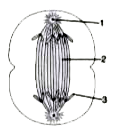
Draw and explain a series of diagrams that illustrate the stages of meiosis I
Step-by-Step Solution for Illustrating the Stages of Meiosis I
Step 1: Prophase I
- Leptotene Stage: Chromatin condenses to form visible chromosomes. Each chromosome consists of two sister chromatids.
- Zygotene Stage: Homologous chromosomes begin to pair up through a process called synapsis, forming bivalents.
- Pachytene Stage: Crossing over occurs between homologous chromosomes, leading to genetic recombination. The centrioles divide and move to opposite poles.
- Diplotene Stage: The bivalents begin to separate slightly, and tetrads (four chromatids) are formed.
- Diakinesis Stage: The nuclear membrane disintegrates, and the chromosomes are fully condensed and ready for alignment.
Diagram for Prophase I: Draw a series of images showing the progression from leptotene to diakinesis, illustrating the changes in chromosome structure and pairing.
---
Step 2: Metaphase I
- In this stage, the tetrads (paired homologous chromosomes) line up at the metaphase plate (the middle of the cell).
- Spindle fibers attach to the centromeres of each homologous chromosome.
Diagram for Metaphase I: Illustrate the tetrads lined up at the center of the cell, with spindle fibers attached to their centromeres.
---
Step 3: Anaphase I
- Early Anaphase I: The homologous chromosomes are pulled apart towards opposite poles of the cell.
- Late Anaphase I: The chromatids are almost at the spindle poles, and the cell membrane begins to constrict.
Diagram for Anaphase I: Show the separation of homologous chromosomes moving towards opposite poles, with a constricting cell membrane.
---
Step 4: Telophase I
- The separated chromosomes reach the poles, and a nuclear membrane starts to form around each set of chromosomes.
- The cell membrane continues to constrict, leading to the formation of two daughter cells.
Diagram for Telophase I: Illustrate the two sets of chromosomes at opposite poles with the nuclear membrane forming around them and the cell membrane constricting.
---
Step 5: Cytokinesis
- This process occurs after telophase I, resulting in two distinct daughter cells, each with the same number of chromatids as the original parent cell.
Diagram for Cytokinesis: Show the final separation of the two daughter cells, indicating that they are identical to the parent cell.
---
Summary of Meiosis I Stages
1. Prophase I: Longest phase with five substages (Leptotene, Zygotene, Pachytene, Diplotene, Diakinesis).
2. Metaphase I: Tetrads align at the metaphase plate.
3. Anaphase I: Homologous chromosomes separate and move to opposite poles.
4. Telophase I: Nuclear membranes form around the separated chromosomes.
5. Cytokinesis: Division of the cytoplasm, resulting in two daughter cells.
- Leptotene Stage: Chromatin condenses to form visible chromosomes. Each chromosome consists of two sister chromatids.
- Zygotene Stage: Homologous chromosomes begin to pair up through a process called synapsis, forming bivalents.
- Pachytene Stage: Crossing over occurs between homologous chromosomes, leading to genetic recombination. The centrioles divide and move to opposite poles.
- Diplotene Stage: The bivalents begin to separate slightly, and tetrads (four chromatids) are formed.
- Diakinesis Stage: The nuclear membrane disintegrates, and the chromosomes are fully condensed and ready for alignment.
- In this stage, the tetrads (paired homologous chromosomes) line up at the metaphase plate (the middle of the cell).
- Spindle fibers attach to the centromeres of each homologous chromosome.
- Early Anaphase I: The homologous chromosomes are pulled apart towards opposite poles of the cell.
- Late Anaphase I: The chromatids are almost at the spindle poles, and the cell membrane begins to constrict.
- The separated chromosomes reach the poles, and a nuclear membrane starts to form around each set of chromosomes.
- The cell membrane continues to constrict, leading to the formation of two daughter cells.
- This process occurs after telophase I, resulting in two distinct daughter cells, each with the same number of chromatids as the original parent cell.
1. Prophase I: Longest phase with five substages (Leptotene, Zygotene, Pachytene, Diplotene, Diakinesis).
2. Metaphase I: Tetrads align at the metaphase plate.
3. Anaphase I: Homologous chromosomes separate and move to opposite poles.
4. Telophase I: Nuclear membranes form around the separated chromosomes.
5. Cytokinesis: Division of the cytoplasm, resulting in two daughter cells.
Topper's Solved these Questions
- CELL CYCLE AND CELL DIVISION BOOK - ICSECHAPTER - CELL CYCLE AND CELL DIVISION EXERCISE - TEST YOUR PROGRESS (HIGHER ORDER THINKING SKILLS QUESTIONS (HOTS))
8 Videos
- CELL CYCLE AND CELL DIVISION BOOK - ICSECHAPTER - CELL CYCLE AND CELL DIVISION EXERCISE - NCERT EXERCISE WITH ANSWER
22 Videos
- CELL CYCLE AND CELL DIVISION BOOK - ICSECHAPTER - CELL CYCLE AND CELL DIVISION EXERCISE - TEST YOUR PROGRESS (VERY SHORT ANSWER TYPE QUESTION)
30 Videos
- CELL : THE UNIT OF LIFE BOOK - ICSECHAPTER - CELL : THE UNIT OF LIFE EXERCISE - Differentiate Between
18 Videos
- CELLULAR RESPIRATION IN PLANTS BOOK - ICSECHAPTER - CELLULAR RESPIRATION IN PLANTS EXERCISE - REVIEW QUESTIONS ( LONG ANSWER TYPE QUESTIONS)
15 Videos
Similar Questions
The longest stage of meiosis is
View SolutionDraw a well labelled diagram showing the stages of the prophase -I of meiosis-I
View SolutionDraw a neat labelled diagram to show the metaphase stage of mitosis in an animal cell having four chromosomes.
View SolutionDraw a well labelled diagram to show the anaphase stage of mitosis in a plant cell having four chromosomes.
View SolutionDraw a well labelled diagram to show various stages of fertilization.
View SolutionDraw suitable diagrams to illustrate the action of (i) concave mirror, and (ii) convex mirror, on a beam of light incident parallel to the principal axis.
View SolutionThe diagram given below represents a stage during cell division. Study the same and then answer the questions that follow :

Name the stage prior to this stage and draw a diagram to represent the same.
View SolutionDraw a labelled ray diagram to illustrate (a) critical angle (b) total internal reflection for a ray of light moving from one medium to another.
View SolutionDraw diagrams to illustrate the positions of fulcrum, load and effort, in each of the following :
(a) A seesaw
(b) A common balance
(c) A nut cracker
(d) Forceps.
View SolutionDraw rough diagrams to illustrate the following: (i) Open curve (ii) Closed curve
View Solution
