Draw the structure of a human heart and label its parts.
Step-by-Step Solution to Draw and Label the Structure of a Human Heart
1. Draw the Outline of the Heart: Start by drawing a rough outline of the heart. The heart is typically shaped like a cone or a slightly tilted pear. Make sure to create a point at the bottom (the apex) and a wider part at the top (the base).
Hint: Remember that the heart is not a perfect shape; it has a slight tilt and is somewhat asymmetrical.
2. Divide the Heart into Chambers: Divide the heart into four sections to represent the four chambers. Draw a vertical line down the middle to separate the left and right sides. Then, draw a horizontal line across the middle to create two upper chambers (auricles) and two lower chambers (ventricles).
Hint: The left side of the heart is responsible for oxygenated blood, while the right side handles deoxygenated blood.
3. Label the Auricles: Label the upper chambers as the "Right Auricle" and "Left Auricle." These are the chambers that receive blood from the body and lungs, respectively.
Hint: Auricles are also known as atria, so you can use either term.
4. Label the Ventricles: Label the lower chambers as the "Right Ventricle" and "Left Ventricle." These chambers pump blood out of the heart to the lungs and the rest of the body, respectively.
Hint: The ventricles are larger and thicker than the auricles because they need to pump blood with more force.
5. Draw and Label the Valves: Indicate the valves between the auricles and ventricles. Label the "Tricuspid Valve" between the right auricle and right ventricle, and the "Mitral Valve" (or Bicuspid Valve) between the left auricle and left ventricle.
Hint: Valves ensure that blood flows in one direction and do not mix oxygenated and deoxygenated blood.
6. Indicate Blood Flow: You can use arrows to show the direction of blood flow. Indicate that deoxygenated blood enters the right auricle, moves to the right ventricle, and is pumped to the lungs. Oxygenated blood enters the left auricle, moves to the left ventricle, and is pumped to the body.
Hint: Use different colors for oxygenated (red) and deoxygenated (blue) blood to make it clearer.
7. Final Touches: Add any additional labels such as "Aorta" (the major artery that carries oxygenated blood from the left ventricle) and "Pulmonary Artery" (which carries deoxygenated blood from the right ventricle to the lungs).
Hint: Make sure all parts are clearly labeled and that the diagram is neat for better understanding.
Summary of the Human Heart Structure
- Right Auricle: Receives deoxygenated blood from the body.
- Left Auricle: Receives oxygenated blood from the lungs.
- Right Ventricle: Pumps deoxygenated blood to the lungs.
- Left Ventricle: Pumps oxygenated blood to the body.
- Tricuspid Valve: Between right auricle and ventricle.
- Mitral Valve: Between left auricle and ventricle.
- Aorta: Carries oxygenated blood to the body.
- Pulmonary Artery: Carries deoxygenated blood to the lungs.
Topper's Solved these Questions
- PRACTICE ASSIGNMENTBOOK - ICSECHAPTER - PRACTICE ASSIGNMENTEXERCISE - PRACTICE ASSIGNMENT 6 (Based on Chapter 6 - Circulatory System) (Fill in the blanks:)
6 Videos
- PRACTICE ASSIGNMENTBOOK - ICSECHAPTER - PRACTICE ASSIGNMENTEXERCISE - PRACTICE ASSIGNMENT PRACTICE ASSIGNMENT 7 (Based on Chapter 7 - Health and Hygiene)
24 Videos
- PRACTICE ASSIGNMENTBOOK - ICSECHAPTER - PRACTICE ASSIGNMENTEXERCISE - PRACTICE ASSIGNMENT 6 (Based on Chapter 6 - Circulatory System)
23 Videos
- HEALTH AND HYGIENE BOOK - ICSECHAPTER - HEALTH AND HYGIENE EXERCISE - Think and answer
3 Videos
- RESPIRATORY SYSTEM BOOK - ICSECHAPTER - RESPIRATORY SYSTEM EXERCISE - TEST YOURSELF (VII. Answer the following quesitons )
3 Videos
Similar Questions
Draw the structure of the human ear and label the following parts: (i) tympanum, (ii) cochlea, (iii) pinna, (iv) malleus, (v) semi-circular canals, (vi) eustachian tube.
View SolutionDraw the microscopic structure of human sperm and relate its different parts with their functions.
View SolutionStudy the diagram given below and answer the questions that follow:

Name the structure shown and label its parts 1 and 2.
View SolutionStudy the diagram given below and answer the questions that follow:
Name the structure shown and label its parts 1 and 2.
View SolutionStudy the diagram given below and answer the questions that follow:

Name the structure shown and label its parts .1. and .2.
View SolutionDraw the well labelled diagram of a mature human sperm and label its parts.
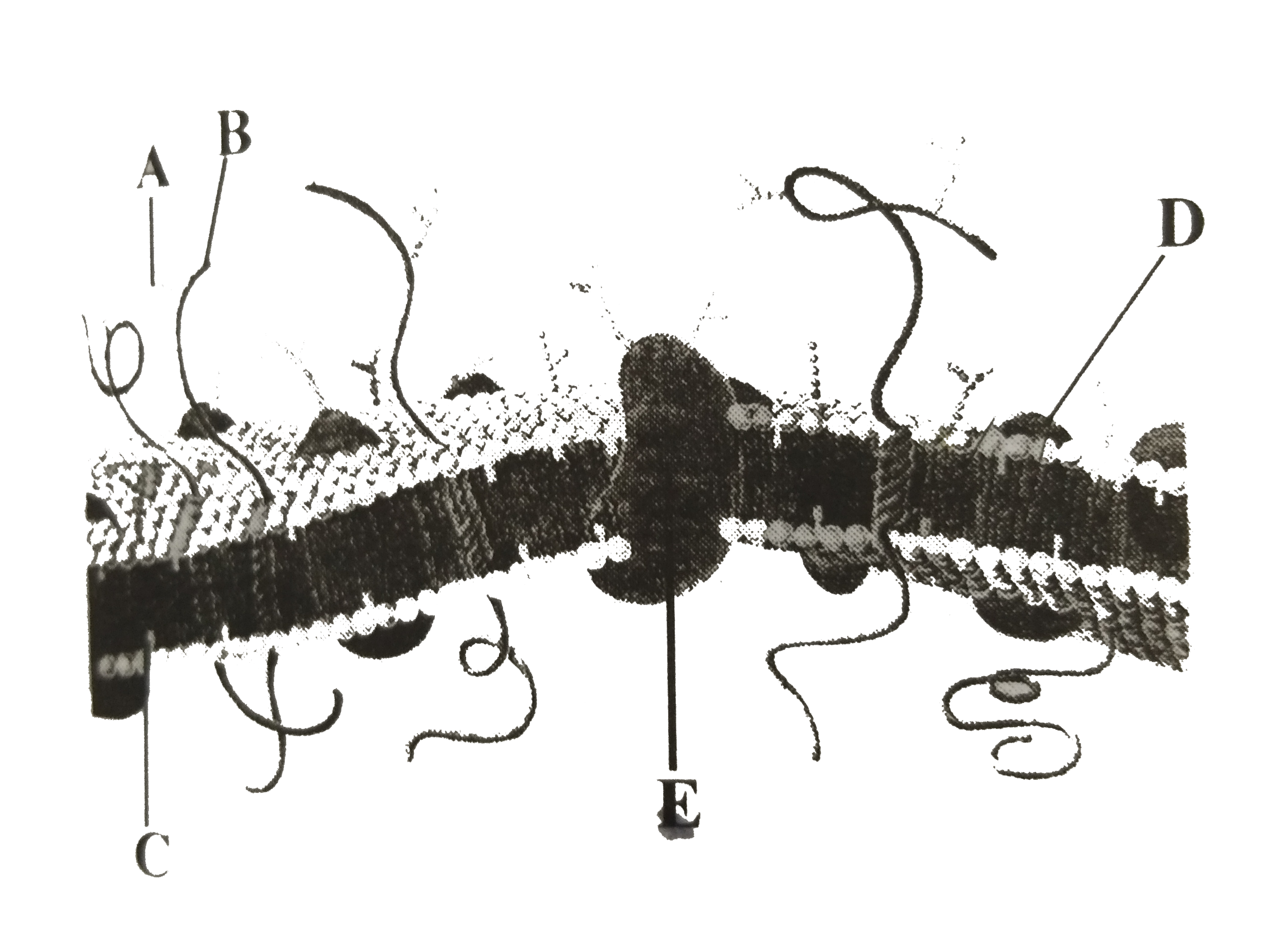
View SolutionThe figure given below show the structure of plasma membrane, with its parts labelled from A to E, Identify the correct :-

View SolutionDraw a well labelled diagram of L.S of ovule showing the structure of embryo sac. Label any six parts.
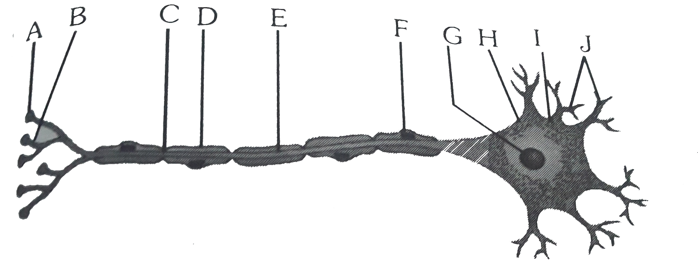
View SolutionGiven below is a structure of a neuron label the parts A to J

View SolutionDraw a schematic diagram of a human sperm and label the cellular components.
View Solution
