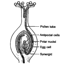
(a) Draw the longitudinal section of a flower showing growth of pollen tube upto the embryo sac.Label the following parts :
(i) Stigma
(ii) Pollen tube
(iii) Integument
(iv) Chalazal end
(v) Nucelllus
(vi) Synergids
(b) What is double fertilisation in Angiosperms? Why is it so called ?
L.S of flower showing growth of pollen tube :

Topper's Solved these Questions
Similar Questions
(a) Draw an L.S. of pistil showing pollen tube entering into the embryo sac. Label the following :
(i) Nucellus (ii) Antipodals
(iii) Synergids (iv) Micropyle
(b) Write the functions of the following :
(i) Synergids (ii) Micropyle
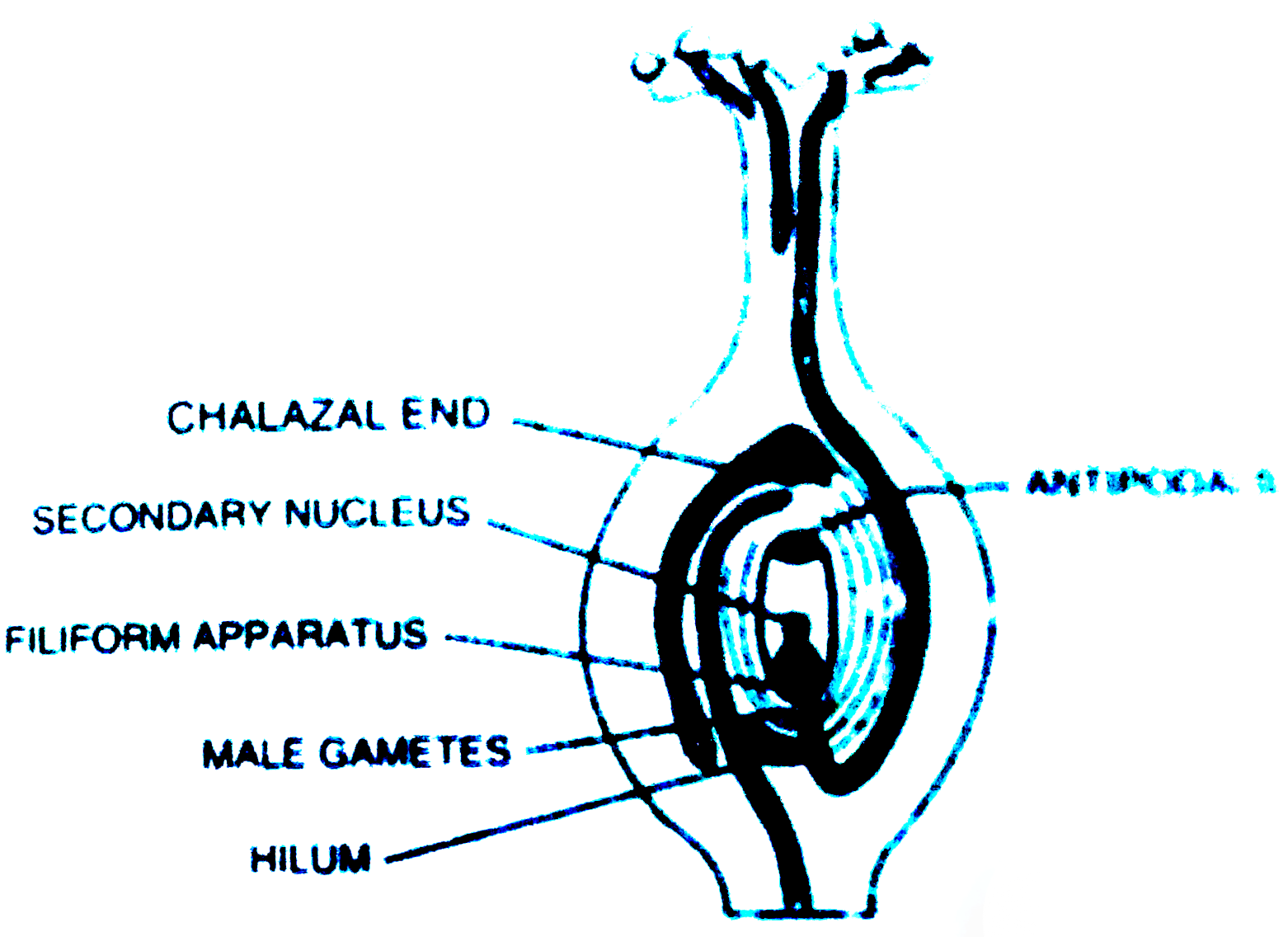
View SolutionDraw a longitudinal section of a post-pollinated pistil showing entry of pollen tube into a mature embryo-sac. Label filiform apparatus, chalazal end, hilum, antipodals male gametes and secondary nucleus .

View SolutionDraw the structure of the human ear and label the following parts: (i) tympanum, (ii) cochlea, (iii) pinna, (iv) malleus, (v) semi-circular canals, (vi) eustachian tube.
View SolutionDraw a longitudinal section of a post-pollinated pistil showing entry of Pollen, tube into a mature embryo-sac. Label filiform apparatus, chalazal end, Hilium, antipodals, male gametes and secondary nucleus.
View SolutionEntry of pollen tube into the embryo sac takes place from a. Chalazal end b. Micropylar end c. Integuments
View Solution(a) Draw a diagram of a mature embryo sac of an angiosperm and label the following parts in it :
(i) Filiform apparatus (ii) Synergids
(iii) Central cell (iv) Egg cell
(v) Polar nuclei (vi) Antipodals
(b) Write the fate of egg cell and polar nuclei after fertilization.
View Solution(a) Draw a diagram of a mature embryo sac of an angiosperm and label the following parts in it :
(i) Filiform apparatus (ii) Synergids
(iii) Central cell (iv) Egg cell
(v) Polar nuclei (vi) Antipodals
(b) Write the fate of egg cell and polar nuclei after fertilization.
View SolutionDraw a longitudinal section of the pistil from a flowering plant, where pollination has occurred. Label the following: (a) Stigma showing germinating pollen grains (b) Style (c) Pollen tube reaching the micropyle of the ovule (d) Embryo sac (e) Components of the egg apparatus
View Solution(a) Draw a diagram of Pistil showing pollen tube growth in angiosperm and label (i) Stigma, (ii) male gametes , (iii) micropyle and (iv) Ovule.
(b) Write the function of micropyle.
View SolutionDraw a diagram of the vertical section of human eye and label the following parts : (i) cornea, (ii) iris, (iii) aqueous humour, (iv) retina, (v) ciliary body, (vi) fovea centralis, (vii) choroid, (viii) pupil, and (ix) lens.
View Solution
