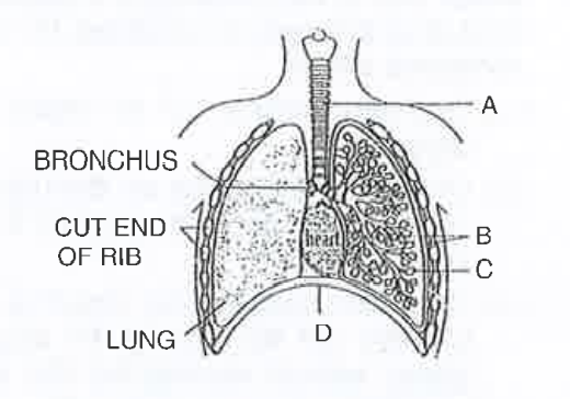
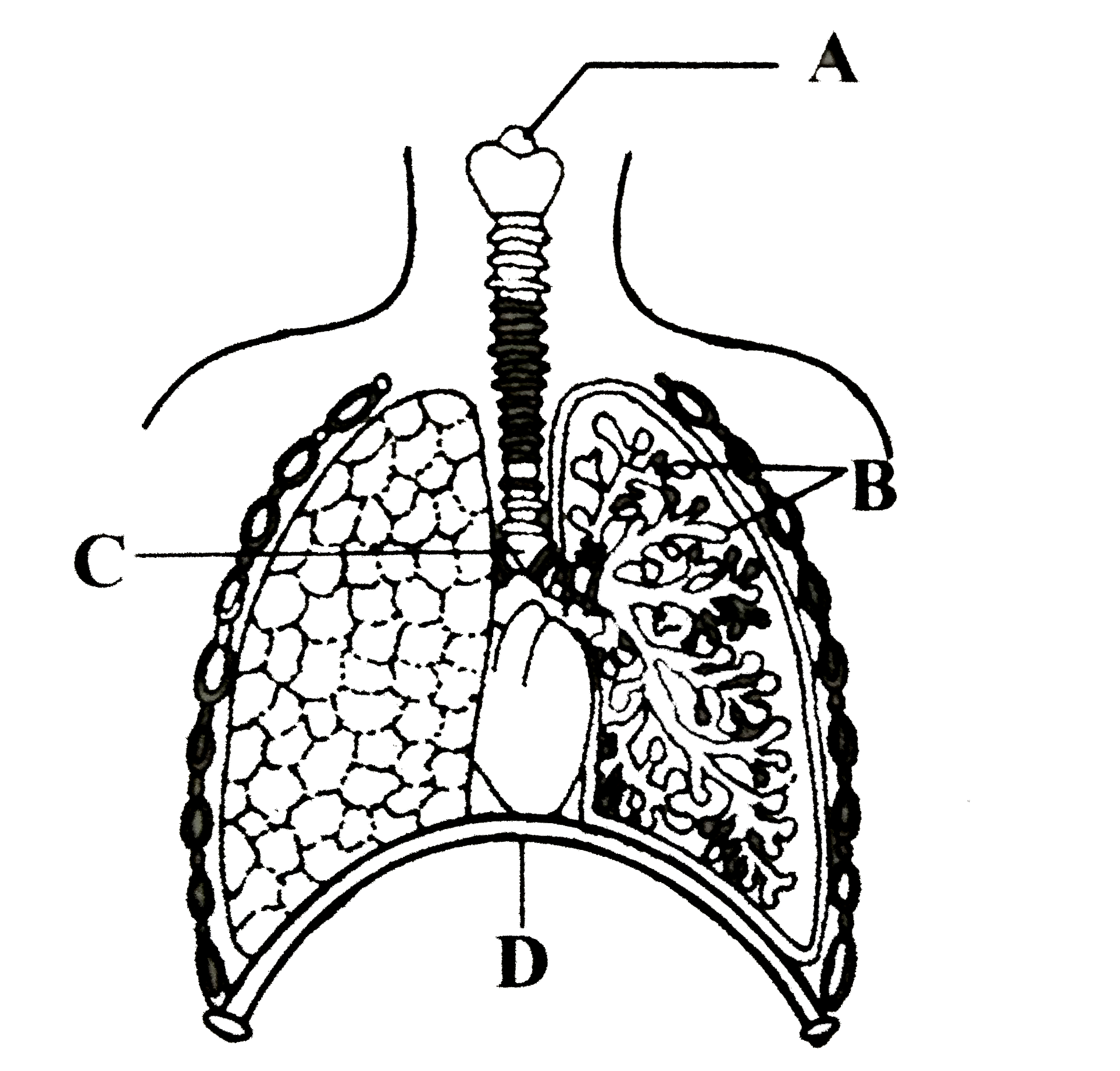
Draw a diagrammatic view of human respiratory system.
To draw a diagrammatic view of the human respiratory system, follow these step-by-step instructions:
1. Start with the Epiglottis and Larynx:
- Draw the epiglottis at the top of your diagram. It is a flap-like structure that covers the trachea during swallowing.
- Below the epiglottis, draw the larynx, which is a box-like structure that contains the vocal cords.
2. Draw the Trachea:
- Below the larynx, draw a long tube representing the trachea.
- Indicate that the trachea is supported by C-shaped cartilaginous rings to keep it open.
3. Divide into Bronchi:
- At the lower end of the trachea, draw a split into two branches, which represent the right and left bronchi. Label them as "Bronchus".
4. Draw Bronchioles:
- From each bronchus, draw smaller branches that represent the bronchioles.
- You can illustrate several levels of branching to show primary, secondary, and tertiary bronchioles.
5. Illustrate Alveoli:
- At the end of the bronchioles, draw clusters of small air sacs to represent the alveoli.
- Label these as "Alveoli", as they are the primary site for gas exchange.
6. Add the Lungs:
- Enclose the bronchi, bronchioles, and alveoli within two large oval shapes to represent the right and left lungs.
- Make sure to label them as "Right Lung" and "Left Lung".
7. Draw the Diaphragm:
- Below the lungs, draw a dome-shaped structure to represent the diaphragm.
- Label it as "Diaphragm", which plays a crucial role in breathing.
8. Include the Heart:
- In the center of the diagram, between the lungs, draw a heart shape to represent the heart.
- Label it as "Heart".
9. Add Pleural Membranes:
- Indicate the pleural membranes surrounding the lungs, and show the pleural fluid between them.
- Label this area as "Pleural Fluid".
Final Diagram:
- Ensure that all the parts are clearly labeled and the diagram is neat and organized.
- Draw the epiglottis at the top of your diagram. It is a flap-like structure that covers the trachea during swallowing.
- Below the epiglottis, draw the larynx, which is a box-like structure that contains the vocal cords.
- Below the larynx, draw a long tube representing the trachea.
- Indicate that the trachea is supported by C-shaped cartilaginous rings to keep it open.
- At the lower end of the trachea, draw a split into two branches, which represent the right and left bronchi. Label them as "Bronchus".
- From each bronchus, draw smaller branches that represent the bronchioles.
- You can illustrate several levels of branching to show primary, secondary, and tertiary bronchioles.
- At the end of the bronchioles, draw clusters of small air sacs to represent the alveoli.
- Label these as "Alveoli", as they are the primary site for gas exchange.
- Enclose the bronchi, bronchioles, and alveoli within two large oval shapes to represent the right and left lungs.
- Make sure to label them as "Right Lung" and "Left Lung".
- Below the lungs, draw a dome-shaped structure to represent the diaphragm.
- Label it as "Diaphragm", which plays a crucial role in breathing.
- In the center of the diagram, between the lungs, draw a heart shape to represent the heart.
- Label it as "Heart".
- Indicate the pleural membranes surrounding the lungs, and show the pleural fluid between them.
- Label this area as "Pleural Fluid".
- Ensure that all the parts are clearly labeled and the diagram is neat and organized.
Topper's Solved these Questions
- SELF ASSESSMENT PAPER 5BOOK - ICSECHAPTER - SELF ASSESSMENT PAPER 5EXERCISE - Part - II SECTION - B (Answer all questions)
11 Videos
- SELF ASSESSMENT PAPER 5BOOK - ICSECHAPTER - SELF ASSESSMENT PAPER 5EXERCISE - Part - II SECTION - C (Answer all questions)
7 Videos
- SELF ASSESSMENT PAPER 5BOOK - ICSECHAPTER - SELF ASSESSMENT PAPER 5EXERCISE - Part - I (Each of the following questions has four choices. Choose the correct option in each case:)
4 Videos
- SELF ASSESSMENT PAPER 4BOOK - ICSECHAPTER - SELF ASSESSMENT PAPER 4EXERCISE - Part - II SECTION - C (Answer all questions)
7 Videos
- SELF ASSESSMENT PAPER-3BOOK - ICSECHAPTER - SELF ASSESSMENT PAPER-3EXERCISE - PART II (SECTION C)
4 Videos
Similar Questions
The figure shows a diagrammatic view of human respiratory system with labels A, B, C and D. Select the option which gives correct identification and main function and/ or characteristic

View SolutionThe figure shows a diagrammatic view of human respiratory system with labels A,B,C and D select the option which gives correct identification and main funciton and / ro characteristic

View Solution
Knowledge Check
The given figure shows the diagrammatic view of human respiratory system. Identify A, B, C and D.
 AA-Epiglottis, B-Alveoli, C-Bronchus, D-DiaphragmBA-Epiglottis, B-Alveoli, C-Bronchioles, D-DiaphragmCA-Soundbox, B-Alveoli, C-Brounchus, D-DiaphragmDA-Larynx, B-Alveoli, C-Brounchioles, D-Diaphragm
a) Draw a labelled diagmmatic view of human male reproductive system.
b) Differentiate between:
i) Vas deferens and vasa efferentia
ii) Spermatogenesis and spermeogenesis
View SolutionDraw a labelled diagram of an human excretory system
View SolutionWhat is the main site of human respiratory system
where exchange of gases occurs?
View SolutionDraw a labelled diagram of the human excretory system.
View SolutionDraw a diagramatic sectional view of human ovary showin stages of oogenesis along with corpus luteum
Where is morula formed in humans? Explain the process of its development from zygote
View SolutionDraw a diagram of human excretory system and label the following :
(i) Urinary bladder
(ii) Left kidney
(iii) Left ureter
View SolutionDraw a diagrammatic sectional view of a human seminiferous tubule, and label sertoli cells, primary spermatocyte, sperm-atogonium and spermatozoa init.
b) Explain the hormonal regulation of the process of spermatogenesis in humans.
View Solution
