Draw a labelled diagram of a synovial joint. Give one example each for a hinge joint, a pivot joint, axial skeleton and appendicular skeleton.
Step-by-Step Solution:
1. Draw a Diagram of a Synovial Joint:
- Start by sketching two bones that meet at a joint.
- Label the following parts:
- Articular Cartilage: A smooth tissue covering the ends of the bones.
- Synovial Membrane: A thin layer lining the joint capsule.
- Joint Cavity: The space between the two bones filled with synovial fluid.
- Synovial Fluid: The lubricating fluid within the joint cavity.
- Articular Capsule: The outer layer that encloses the joint.
2. Examples of Different Joints:
- Hinge Joint: Example - Elbow Joint. This joint allows movement in one plane, like opening and closing a door.
- Pivot Joint: Example - Atlantoaxial Joint (between the first and second cervical vertebrae). This joint allows rotational movement.
- Axial Skeleton: Example - Skull. The axial skeleton consists of the bones of the head and trunk.
- Appendicular Skeleton: Example - Femur (thigh bone). The appendicular skeleton includes the bones of the limbs and girdles.
- Start by sketching two bones that meet at a joint.
- Label the following parts:
- Articular Cartilage: A smooth tissue covering the ends of the bones.
- Synovial Membrane: A thin layer lining the joint capsule.
- Joint Cavity: The space between the two bones filled with synovial fluid.
- Synovial Fluid: The lubricating fluid within the joint cavity.
- Articular Capsule: The outer layer that encloses the joint.
- Hinge Joint: Example - Elbow Joint. This joint allows movement in one plane, like opening and closing a door.
- Pivot Joint: Example - Atlantoaxial Joint (between the first and second cervical vertebrae). This joint allows rotational movement.
- Axial Skeleton: Example - Skull. The axial skeleton consists of the bones of the head and trunk.
- Appendicular Skeleton: Example - Femur (thigh bone). The appendicular skeleton includes the bones of the limbs and girdles.
Topper's Solved these Questions
- LOCOMOTIONA ND MOVEMENT BOOK - ICSECHAPTER - LOCOMOTIONA ND MOVEMENT EXERCISE - TEST YOUR PROGRESS ( HIGHER ORDER THINKING SKILLS QUESTIONS (HOTS))
9 Videos
- LOCOMOTIONA ND MOVEMENT BOOK - ICSECHAPTER - LOCOMOTIONA ND MOVEMENT EXERCISE - NCERT EXERCISES WITH ANSWER
19 Videos
- LOCOMOTIONA ND MOVEMENT BOOK - ICSECHAPTER - LOCOMOTIONA ND MOVEMENT EXERCISE - TEST YOUR PROGRESS ( VERY SHORT ANSWER TYPE QUESTIONS )
73 Videos
- LOCOMOTION AND MOVEMENTBOOK - ICSECHAPTER - LOCOMOTION AND MOVEMENTEXERCISE - LONG ANSWER TYPE QUESTIONS
14 Videos
- MINERAL NUTRITION BOOK - ICSECHAPTER - MINERAL NUTRITION EXERCISE - NCERT EXERCISE WITH ANSWER (DIFFERNTIATE BETWEEN )
9 Videos
Similar Questions
Draw a labelled diagram of a class II lever. Give one example of such a lever.
View SolutionWhat is a joint? Give one example of an immovable joints.
View SolutionGive one example each of fibrous joint and cartilaginous joint.
View SolutionGive and example of saddle joint.
View SolutionGive an example for each of the following:
A synovial joint
View SolutionDraw a labelled sketch of a class III lever. Give one example of this kind of lever.
View SolutionThe example of pivot joint is
View SolutionWhich of the following is not the example of synovial joint?
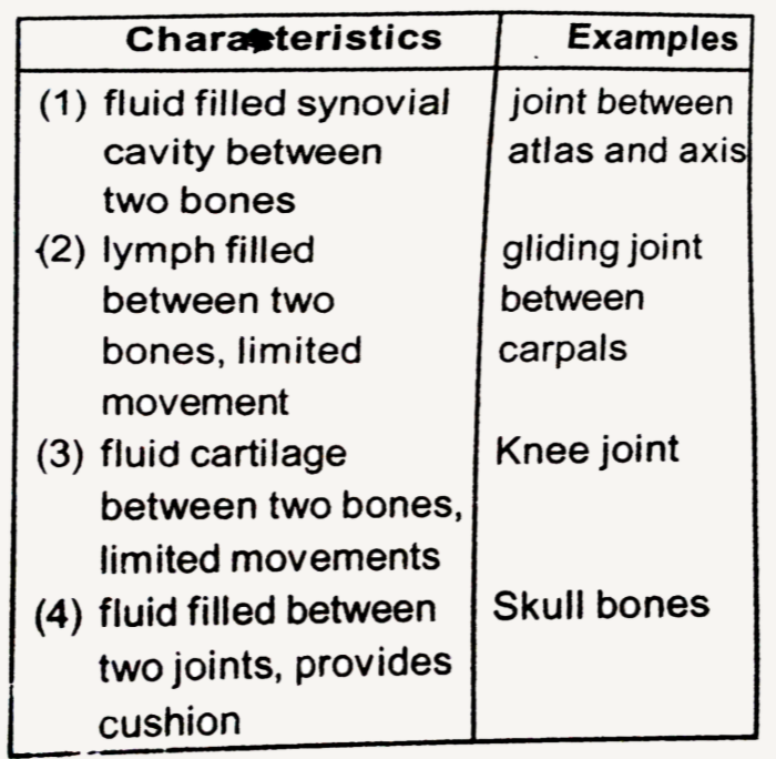
View SolutionThe characteristics and an example of a synovial joint in humans is

View SolutionWhich one of the following is not a synovial joint
View Solution
