Ventricular contraction is in command of :−
SA node
AV node
Purkinje fibers
Papillary muscles
The correct Answer is:A
Step-by-Step Solution:
1. Understanding the Heart's Conduction System: The heart has a specialized conduction system that controls the rhythm and timing of contractions. The main components include the Sinoatrial (SA) node, Atrioventricular (AV) node, Bundle of His, and Purkinje fibers.
2. Role of the SA Node: The SA node, located in the right atrium, is known as the natural pacemaker of the heart. It generates electrical impulses that initiate the contraction of the atria.
3. Transmission to the AV Node: After the SA node fires, the electrical signal travels to the AV node, which is located in the inter-atrial septum. The AV node serves as a gatekeeper that delays the impulse slightly to ensure that the atria have time to contract and fill the ventricles with blood.
4. Conduction through the Bundle of His: Following the delay at the AV node, the impulse travels down the Bundle of His, which splits into the left and right bundle branches that extend into the ventricles.
5. Activation of Purkinje Fibers: The electrical signal is then conducted to the Purkinje fibers, which spread throughout the ventricles. This is crucial as the Purkinje fibers ensure that the contraction of the ventricles is synchronized and occurs effectively.
6. Conclusion: The Purkinje fibers are responsible for commanding the ventricular contraction, making them the correct answer to the question. Other options, such as the SA node, AV node, and papillary muscles, do not directly control ventricular contraction.
Final Answer:
Ventricular contraction is in command of Purkinje fibers.
---
Ventricular contraction is in command of Purkinje fibers.
Topper's Solved these Questions
- BODY FLUIDS AND CIRCULATIONS BOOK - CENGAGE BIOLOGY ENGLISHCHAPTER - BODY FLUIDS AND CIRCULATIONS EXERCISE - Exercise (Assertion - Reasoning Questions)
15 Videos
- BODY FLUIDS AND CIRCULATIONS BOOK - CENGAGE BIOLOGY ENGLISHCHAPTER - BODY FLUIDS AND CIRCULATIONS EXERCISE - ARCHIVES (Choose the correct option)
30 Videos
- BIOTECHNOLOGY:PRINCIPLES AND PROCESSESBOOK - CENGAGE BIOLOGY ENGLISHCHAPTER - BIOTECHNOLOGY:PRINCIPLES AND PROCESSESEXERCISE - ARCHIVES (Choose the correct option)
33 Videos
- ECOSYSTEMBOOK - CENGAGE BIOLOGY ENGLISHCHAPTER - ECOSYSTEMEXERCISE - ARCHIVES (CHOOSE THE CORRECT OPTION)
41 Videos
Similar Questions
Knowledge Check
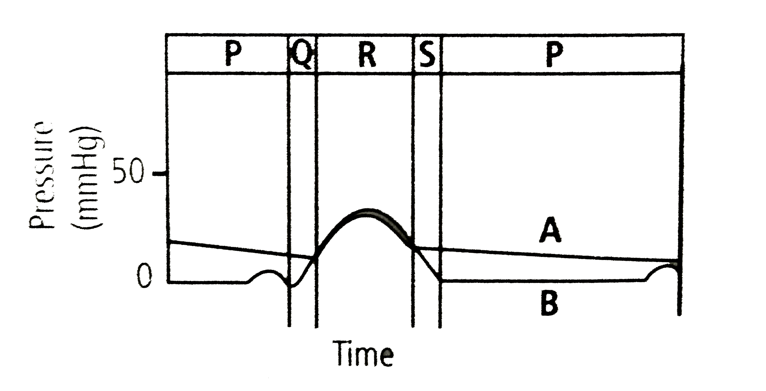
Refer to the given figure in which A refers to pulmonary artery pressure and B refers to right ventricular pressure. Identify P,Q,R and S in the figure and match with the list (i-iv) given below
(i) Isovolumetric ventricular contraction
(ii) Ventricular ejection
(iii) Isovolumetric ventricular relaxation
(iv) Ventricular filling
 AP-(iv),Q-(iii),R-(ii),S-(i)BP-(ii),Q-(iii),R-(i),S-(iv)CP-(iv),Q-(i),R-(ii),S-(iii)DP-(i),Q-(ii),R-(iii),S-(iv)
A sustained contraction is called
View SolutionDuring muscle contraction
View SolutionDuring muscle contraction
View SolutionDuring muscle contraction
View SolutionInvoluntary muscular contraction is called
View SolutionDuring muscle contraction
View SolutionPresence of which of the following hormones can increase the strength of ventricular contraction, br a. Epinephrine, br b. Acetylcholine, br c. Nor adrenaline, br d. Catecholamines
View Solution
