Given figures (IandII) show two specialised connective tissues. Identify the figures I,II and the parts labelled as A and B
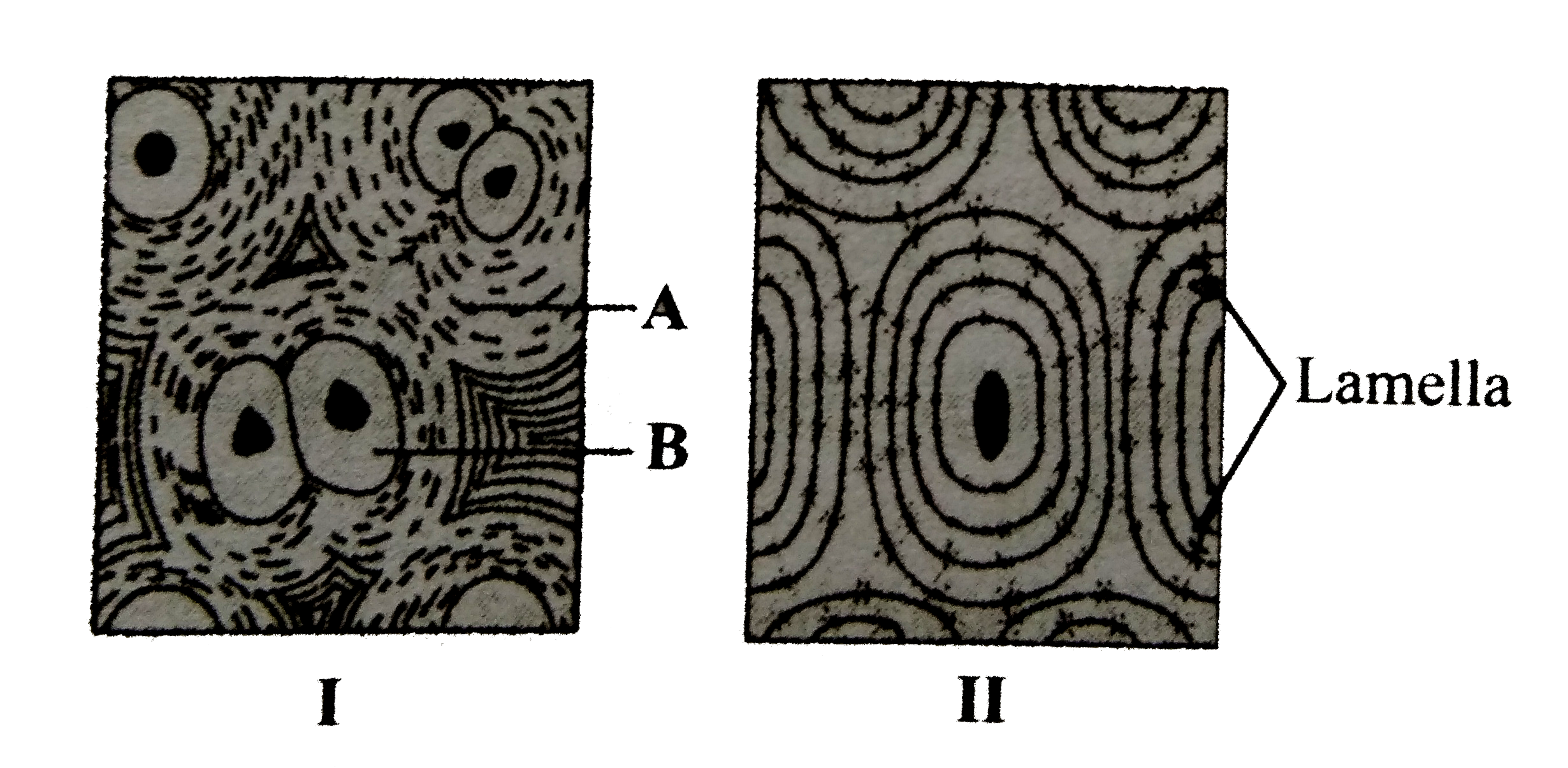
I - Bone, II - Cartilage, C - Collagen fibers, D - Osteoblast
I - Cartilage, II - Bone, A - Collagen fibers, B- Chondroclast
I - Bone, II - Cartilage, C- Elastin fibres, D - Osteoblast
I - Cartilage, II - Bone, A - Collagen fibers, B-Chondrocyte
The correct Answer is:D
Step by step video & image solution for Given figures (I and II) show two specialised connective tissues. Identify the figures I,II and the parts labelled as A and B by Biology experts to help you in doubts & scoring excellent marks in Class 12 exams.
Updated on:21/07/2023Topper's Solved these Questions
- STRUCTURAL ORGANISATION IN ANIMALS BOOK - VMC MODULES ENGLISHCHAPTER - STRUCTURAL ORGANISATION IN ANIMALS EXERCISE - EFFICIENT
50 Videos
- STRUCTURAL ORGANISATION IN ANIMALS BOOK - VMC MODULES ENGLISHCHAPTER - STRUCTURAL ORGANISATION IN ANIMALS EXERCISE - IMPECCABLE
50 Videos
- STRUCTURAL ORGANISATION IN ANIMALS BOOK - VMC MODULES ENGLISHCHAPTER - STRUCTURAL ORGANISATION IN ANIMALS EXERCISE - In-Chapter Exercise-G
10 Videos
- STRATEGIES FOR ENHANCEMENT IN FOOD PRODUCTION [PLANT BREEDING]BOOK - VMC MODULES ENGLISHCHAPTER - STRATEGIES FOR ENHANCEMENT IN FOOD PRODUCTION [PLANT BREEDING]EXERCISE - In Chapter Exercise - B
10 Videos
- THE LIVING WORLD BOOK - VMC MODULES ENGLISHCHAPTER - THE LIVING WORLD EXERCISE - CHAPTER EXERCISE-D
9 Videos
Similar Questions
Knowledge Check
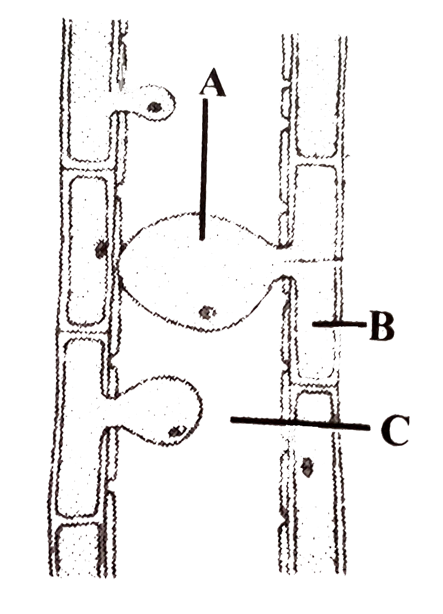
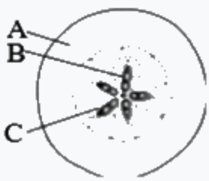
Identify the given figure and select the correct labels for A,B and CAABCCalloseXylem parenchymaXylem vesselBABCCallosePhloem parenchymaPhloem vesselCABCTylosisXylem parenchymaXylem vesselDABCTylosisPhleom parenchymaPhloem vesselRefer to the given figure and identify the parts labelled P,Q,R and S.
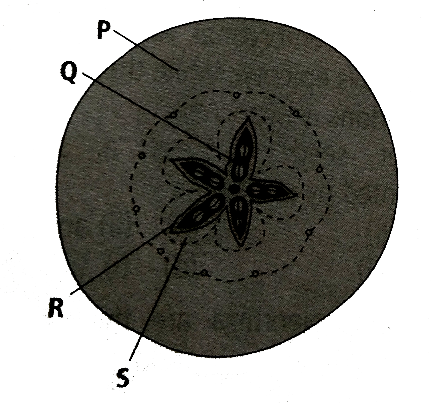 APQRSSeedThalamusMesocarpEndocarpBPQRSThalamusSeedEndocarpMesocarpCPQRSSeedThalamusEndocarpMesocarpDPQRSPericapSeedMesocarpEndocarp
Given is the diagrammatic sketch of a certain type of connective tissue
Identify the parts labelled as A,B,C and D and select the correct option.
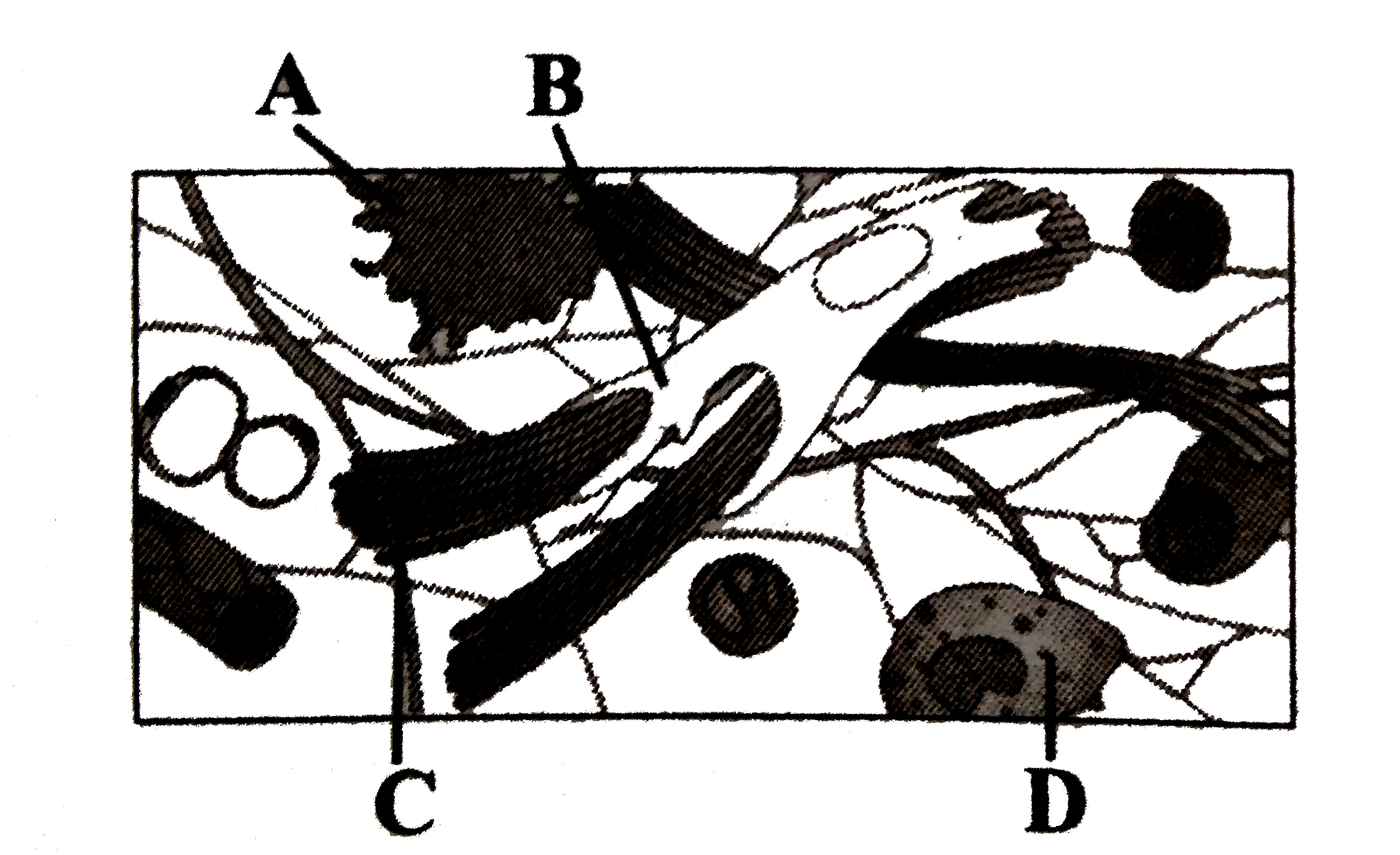 A A B C D Macrophage Fibroblast Collagen fibres Mast cellB A B C D Mast cell Macrophage Fibroblast Collagen fibreC A B C D Macrophage Collagen fibre Fibroblast Mast cellD A B C D Mast cell Collagen fibre Fibroblast Macrophage
In the given figure label the parts

View SolutionThe diagram given below shows the supportive connective tissue.

Identify the tissues A and B.
View SolutionIdentify the given figure with correct labelling :
View SolutionStudy the given figure and identify the cells labelled as A, B, C and D
View SolutionGiven is the diagrammatic sketch of a certain type of connective tissue
Identify the parts labelled as A,B,C and D and select the correct option.

View Solution
