A picture of dividing Drosophila cell through a microscope show that the cell contains 8 chromosomes, each consisting of two chromatids. The picture must have been taken during.
Mitotic metaphase
Mitotic Anaphase
Meiotic Anaphase II
Meiotic Telophase II
The correct Answer is:A
To determine when the picture of a dividing Drosophila cell with 8 chromosomes, each consisting of two chromatids, was taken, we can follow these steps:
1. Understand Chromosome Structure: Each chromosome in the picture has two chromatids. This indicates that the chromosomes have already been replicated. In a typical cell cycle, chromosomes are replicated during the S phase of interphase.
2. Identify the Total Number of Chromosomes: The picture shows a total of 8 chromosomes. Since each chromosome consists of two chromatids, this means that the cell is in a stage of division where the chromatids are still joined together.
3. Determine the Stage of Cell Division: The cell division process includes several stages: prophase, metaphase, anaphase, and telophase. During metaphase, chromosomes are aligned at the metaphase plate and are still in the form of sister chromatids.
4. Eliminate Other Stages:
- Anaphase: In this stage, sister chromatids are pulled apart towards opposite poles of the cell. Therefore, if the cell had reached anaphase, we would not see chromosomes with two chromatids.
- Telophase: At this stage, the chromatids have already separated and are being enclosed in new nuclear membranes. Thus, we would not see paired chromatids.
- Prophase: While chromosomes are visible, they are not yet aligned at the metaphase plate.
5. Conclude the Correct Stage: Since the picture shows chromosomes that are still paired as sister chromatids and aligned, it is clear that the picture was taken during metaphase. This is the only stage where chromosomes are arranged on the metaphase plate and consist of two chromatids.
Final Answer:
The picture must have been taken during metaphase.
---
- Anaphase: In this stage, sister chromatids are pulled apart towards opposite poles of the cell. Therefore, if the cell had reached anaphase, we would not see chromosomes with two chromatids.
- Telophase: At this stage, the chromatids have already separated and are being enclosed in new nuclear membranes. Thus, we would not see paired chromatids.
- Prophase: While chromosomes are visible, they are not yet aligned at the metaphase plate.
The picture must have been taken during metaphase.
Similar Questions
Prior to meiosis, DNA replication occurs, so each chromosome contains two sister chromatids that are identical to the original chromosome. Meiosis is divided into two nuclear divisions: meiosis I and meiosis II. Each of these nuclear divisions shares many aspects of mitosis and can be divided into the same phases: prophase, metaphase, anaphase, and telophase, however, between the two divisions, DNA replication does not occur. Through this process, one diploid cell will divide into four haploid cells. During meiosis I. the pairs of homologous chromosomes are separated from each other. During prophase I, the homologous chromosomes line up together. During this time, crossing over canoccur(theexchangeofDNA betweenhomologous chromosomes). Crossing-over increases the new allele combinations in the gametes. Without crossing-over, the offspring would always inherit all of the many alleles on one of the homologous chromosomes. (An allele is an alternative form of a gene which is located at a specific position on a specific chromosome.) Because of crossing-over, the alleles on the homologous chromosomes can be scrambled to pass on unique combinations of alleles on the chromosome. Also, during prophase I. the spindle forms and the chromosomes condense as they coil up tightly. The spindle has the same function as in mitosis.
How many haploid cells will result from one diploid cell after it has undergone meiosis?View SolutionPrior to meiosis, DNA replication occurs, so each chromosome contains two sister chromatids that are identical to the original chromosome. Meiosis is divided into two nuclear divisions: meiosis I and meiosis II. Each of these nuclear divisions shares many aspects of mitosis and can be divided into the same phases: prophase, metaphase, anaphase, and telophase, however, between the two divisions, DNA replication does not occur. Through this process, one diploid cell will divide into four haploid cells. During meiosis I. the pairs of homologous chromosomes are separated from each other. During prophase I, the homologous chromosomes line up together. During this time, crossing over canoccur(theexchangeofDNA betweenhomologous chromosomes). Crossing-over increases the new allele combinations in the gametes. Without crossing-over, the offspring would always inherit all of the many alleles on one of the homologous chromosomes. (An allele is an alternative form of a gene which is located at a specific position on a specific chromosome.) Because of crossing-over, the alleles on the homologous chromosomes can be scrambled to pass on unique combinations of alleles on the chromosome. Also, during prophase I. the spindle forms and the chromosomes condense as they coil up tightly. The spindle has the same function as in mitosis.
During prophase I, a unique process occurs. Provide the name and brief definition of this process.View Solution
Knowledge Check
- Question 1 - Select One
Assertion :- During M phase of karyokinesis of mitosis each chromosomes is made up of two chromatids.
Reason:- Before M phase DNA content have already been doubled during S-phase.AIf both Assertion & Reason are True & the Reason is a correct explanation of the Assertion.BIf both Assertion & Reason are True but Reason is not a correct explanation of the AssertionCIf Assertion is True but the Reason is FalseDIf both Assertion & Reason are false. - Question 2 - Select One
At what phase of meiosis there are two cells, each with separated siter chromatids that have been moved to opposite spindle poles?
AAnaphase IIBAnaphase ICTelophase IIDTelophase I - Question 3 - Select One
Choose the correct statements with respect to the completion of prophase
I. Chromosomal material condenses to from compact mitotic chromosomes. Chromosomes are seen to be composed of two chromatids attached together at the centromere.
II. Initiation of the assembly of mitotic spindle, the microtubules, the proteinaceous components of the cell cytoplasm
III. Cells at the end of prophase, when viewed under the microscope, do not show golgi complexes, endoplasmic reticulum, nucleolus and the nuclear envelopeAI onlyBII onlyCI and IIDI, II and III
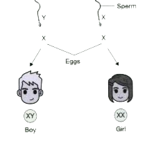
The life of a human being begins as a single celled zygote. The information presents in the chromosomes in this cell is what determines the characteristics of this human being. Each cell in the body of a male contains one X chro mosome and one Y chromosome (XY).
Read the passage and try to answer the questions

What chromosomes do females have?View SolutionPrior to meiosis, DNA replication occurs, so each chromosome contains two sister chromatids that are identical to the original chromosome. Meiosis is divided into two nuclear divisions: meiosis I and meiosis II. Each of these nuclear divisions shares many aspects of mitosis and can be divided into the same phases: prophase, metaphase, anaphase, and telophase, however, between the two divisions, DNA replication does not occur. Through this process, one diploid cell will divide into four haploid cells. During meiosis I. the pairs of homologous chromosomes are separated from each other. During prophase I, the homologous chromosomes line up together. During this time, crossing over canoccur(theexchangeofDNA betweenhomologous chromosomes). Crossing-over increases the new allele combinations in the gametes. Without crossing-over, the offspring would always inherit all of the many alleles on one of the homologous chromosomes. (An allele is an alternative form of a gene which is located at a specific position on a specific chromosome.) Because of crossing-over, the alleles on the homologous chromosomes can be scrambled to pass on unique combinations of alleles on the chromosome. Also, during prophase I. the spindle forms and the chromosomes condense as they coil up tightly. The spindle has the same function as in mitosis.
Briefly describe the similarities between the meiosis and mitosis processes.View SolutionGiven here are three statements about cell cycle.
I. Synthesis of histones and replication of DNA both occur during S-phase of inter phase.
II. After S-phase, each cell has double the number of chromosomes.
III. Separation of sister chromatids takes place during G2− phase
Which of the given statements is/are true about cell cycle?View SolutionGyandromorphs develop in Drosophila when the two cells in the two-called proembryo will have one of the following chromosomal sets
View SolutionA Mitosis produces two identical daughter cell.
B Meiosis introduces variation due to gene exchange or recombination.
C During anaphase chromosome have two chromatids.
D Telophase I may be absent.
Choose the option with the correct statements.View Solution

