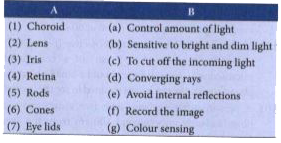
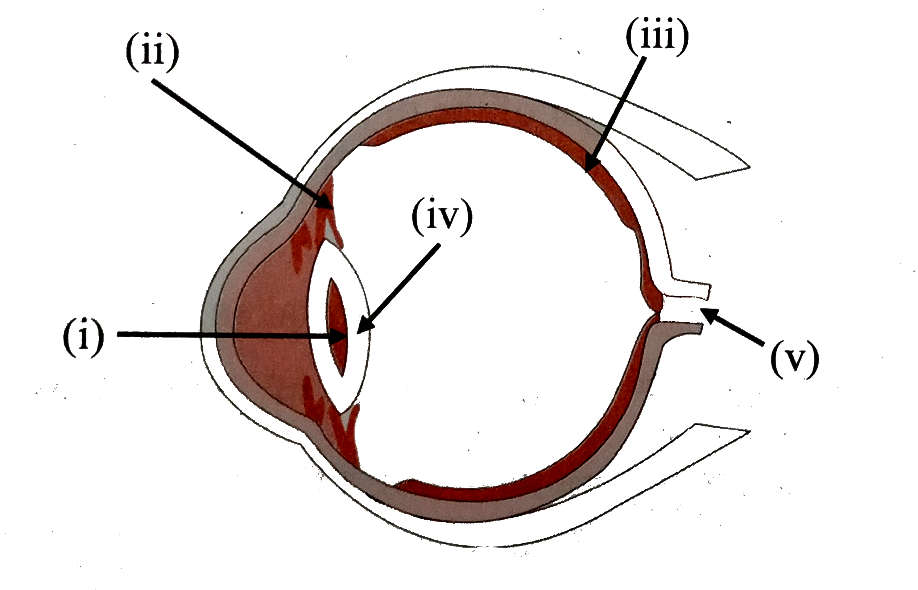
Draw a labelled sketch of the human eye. Explain the functions of each part.
Step-by-Step Text Solution:
1. Draw the Sketch of the Human Eye:
- Begin by sketching the outline of the human eye. Include the following parts: cornea, pupil, iris, lens, retina, ciliary muscles, aqueous humor, optic nerve, and blind spot.
2. Label the Parts:
- Clearly label each part of the eye in your sketch:
- Cornea
- Pupil
- Iris
- Lens
- Retina
- Ciliary Muscles
- Aqueous Humor
- Optic Nerve
- Blind Spot
3. Explain the Functions of Each Part:
- Cornea: The outer transparent layer that allows light to enter the eye and provides a slight refraction of light.
- Pupil: The small opening in the center of the iris that controls the amount of light entering the eye.
- Iris: The colored part of the eye that surrounds the pupil and adjusts its size to regulate light intake.
- Lens: A jelly-like convex lens that focuses light onto the retina, allowing for clear images.
- Retina: The inner layer that acts as a screen where light-sensitive cells detect light and form images.
- Ciliary Muscles: Muscles that adjust the shape of the lens to change its focal length, enabling the eye to focus on objects at various distances.
- Aqueous Humor: The fluid in the front part of the eye that helps maintain intraocular pressure and provides nutrients to the eye tissues.
- Optic Nerve: The nerve that transmits visual information from the retina to the brain for processing.
- Blind Spot: The area on the retina where there are no light-sensitive cells, resulting in a lack of vision at that point.
- Begin by sketching the outline of the human eye. Include the following parts: cornea, pupil, iris, lens, retina, ciliary muscles, aqueous humor, optic nerve, and blind spot.
- Clearly label each part of the eye in your sketch:
- Cornea
- Pupil
- Iris
- Lens
- Retina
- Ciliary Muscles
- Aqueous Humor
- Optic Nerve
- Blind Spot
- Cornea: The outer transparent layer that allows light to enter the eye and provides a slight refraction of light.
- Pupil: The small opening in the center of the iris that controls the amount of light entering the eye.
- Iris: The colored part of the eye that surrounds the pupil and adjusts its size to regulate light intake.
- Lens: A jelly-like convex lens that focuses light onto the retina, allowing for clear images.
- Retina: The inner layer that acts as a screen where light-sensitive cells detect light and form images.
- Ciliary Muscles: Muscles that adjust the shape of the lens to change its focal length, enabling the eye to focus on objects at various distances.
- Aqueous Humor: The fluid in the front part of the eye that helps maintain intraocular pressure and provides nutrients to the eye tissues.
- Optic Nerve: The nerve that transmits visual information from the retina to the brain for processing.
- Blind Spot: The area on the retina where there are no light-sensitive cells, resulting in a lack of vision at that point.
Topper's Solved these Questions
Similar Questions
Draw a labelled figure of the V.S. of human eye.
View SolutionDraw a labelled sketch of wastewater treatment plant .
View SolutionDraw a labelled diagram of eye .
View SolutionDraw a labelled diagram of human heart.
View SolutionDraw a well labelled diagrams of human egg
View SolutionMatch the parts of the human eye with its function .

View SolutionDraw a neat labelled sketch of a replicating fork of DNA.
View SolutionDraw a labelled sketch of the section of a mature pollen grain.
View SolutionLabel the parts of the following diagram of the human eye and state the functions of those parts.

View SolutionDraw a well labelled diagram of human sperm.
View Solution