Myofibrils show dark and light bands is
Cardiac muscles
Unstriped muscles
Striped muscles
Both A and C.
The correct Answer is:D
Step-by-Step Solution:
1. Identify the Types of Muscles: The question asks about myofibrils showing dark and light bands in different types of muscles. The three types of muscles to consider are:
- Cardiac Muscle
- Smooth Muscle (Unstriated Muscle)
- Skeletal Muscle (Striated Muscle)
2. Understand Myofibril Structure: Myofibrils are the contractile elements in muscle fibers. They are composed of repeating units called sarcomeres, which contain thick (myosin) and thin (actin) filaments.
3. Recognize the Presence of Striations:
- Skeletal Muscle: This type of muscle is striated, meaning it has alternating dark (A bands) and light (I bands) regions due to the arrangement of myosin and actin filaments.
- Cardiac Muscle: Cardiac muscle is also striated and contains dark and light bands similar to skeletal muscle, due to the presence of myofibrils.
- Smooth Muscle: This type of muscle is unstriated and does not show distinct dark and light bands.
4. Conclusion: Based on the above understanding, both cardiac muscle and skeletal muscle show dark and light bands due to the presence of myofibrils. Smooth muscle does not show these bands.
5. Final Answer: Therefore, the correct answer to the question is that myofibrils show dark and light bands in both cardiac muscles (A) and skeletal muscles (C).
- Cardiac Muscle
- Smooth Muscle (Unstriated Muscle)
- Skeletal Muscle (Striated Muscle)
- Skeletal Muscle: This type of muscle is striated, meaning it has alternating dark (A bands) and light (I bands) regions due to the arrangement of myosin and actin filaments.
- Cardiac Muscle: Cardiac muscle is also striated and contains dark and light bands similar to skeletal muscle, due to the presence of myofibrils.
- Smooth Muscle: This type of muscle is unstriated and does not show distinct dark and light bands.
Topper's Solved these Questions
- STRUCTURAL ORGANISATION IN ANIMALS BOOK - DINESH PUBLICATION ENGLISHCHAPTER - STRUCTURAL ORGANISATION IN ANIMALS EXERCISE - Check your grasp
10 Videos
- STEM BOOK - DINESH PUBLICATION ENGLISHCHAPTER - STEM EXERCISE - MULTIPLE CHOICE QUESTIONS
134 Videos
- STRUCTURE OF THE CELLBOOK - DINESH PUBLICATION ENGLISHCHAPTER - STRUCTURE OF THE CELLEXERCISE - Check your Grasp
17 Videos
Similar Questions
Myofibrils are
View SolutionRead the statement regarding myofibril and choose the correct option
1. Each myofibril has alternate dark and light bands.
2. In the centre of each 'A' band is an elastic fibre called 'Z' -line 3. 'A' and 'I' bands are arranged alternately throughout the length of myofibril
4. Sarcomere is the functional unit of contraction
5. Central part of thick filament not overlapped by a thin filament is the 'M' line
View Solution
Knowledge Check
Dark bands are
AA-bandBB-bandCI-bandDZiline.
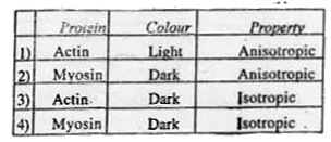
Skeletal muscle appears striated due to the presence of two characteristic proteins in alternating dark and light bands. Which of the following is a correct match of the protein with its light refractive property and colour?

View SolutionRefer to the characteristics given below and identify how many are true for a skeletal muscle ?
Uninucleated , branched , dark and light band , actin and myosin filaments , spindle - shaped cells
View SolutionPoints which are correct with respect to cardiac muscle ?
(i) Have dark and light bands
(ii) Involuntory in nature
(iii) Similar to smooth muscle in structure
(iv) Muscle fibres are branched, cylindrical
(v) get fatigued after a long period.
View SolutionThe distance or length of the myofibril between two adjacent z-bands is called
View SolutionA-band and l-band
View SolutionIn a myofibril, each I band is bisected by
View SolutionIn a myofibril , the band which contains only actin filament is also known as
View Solution
