Skeletal muscles have a striped appearance under the microscope and hence called____mucles.
The correct Answer is:Striated
Step-by-Step Solution:
1. Understanding Skeletal Muscles: Skeletal muscles are the muscles that are attached to the skeleton and are responsible for voluntary movements of the body. They are found in various parts of the body, including the limbs.
2. Microscopic Appearance: When observed under a microscope, skeletal muscles exhibit a distinctive striped or banded appearance. This characteristic is crucial for identifying these muscles.
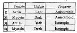
3. Terminology: The striped appearance of skeletal muscles is due to the arrangement of protein filaments within the muscle fibers. These filaments are primarily actin (thin filaments) and myosin (thick filaments).
4. Striated Muscles: Because of their striped appearance, skeletal muscles are referred to as "striated muscles." The term "striated" comes from the Latin word "stria," meaning "stripe" or "furrow."
5. Conclusion: Therefore, the answer to the question is that skeletal muscles are called "striated muscles" due to their striped appearance under the microscope.
Final Answer: Skeletal muscles have a striped appearance under the microscope and hence called striated muscles.
---
Topper's Solved these Questions
Similar Questions
Knowledge Check
The structure in chromatin seen as 'beads-on' string' when viewed under electron microscope are called
AnucleotidesBnuclesidesChistone octamerDnucleosomes.A scientist was studying the production of a protein that was released by an animal cell into a culture medium, she found that the protein only appeared in the culture medium after the added a few drops of a hormone to the cell. Before adding the hormone, she labelled the protein inside the cell with a fluorescent dye and looked at the cell under the lgiht microscope. The dye was seen in flattened sheets and tube. like structures throughout the cell, and in stacks of flattened sac-like structures. after adding the hormone, the dye was aslo seen as small dots clustered against the cell membrane, Which statement most likely explains these observations?
AThe hormone stimulates protein synthesis in the cell vacuole, the protein is then passed to the Golgi apparatus, and eventually passess through the cell membrane by passive diffusion.BThe hormone triggers the synthesis of the protein in the endoplasmic reticulum and it is then secreted outside of the cell via channel proteins in the cell membraneCThe protein is made in the endoplasmic reticulum, is passed to the Golgi appartus and is secreted through hormone-stimulated exocytosis.DThe protein is made in the Golgi apparatus, is passed to the endoplasmic recticulum and is secreted through hormone-stimulated pinocytosis.
How many of the following features belong to skeletal muscles?
I. Striped appearance
II. Voluntary nature
III. Not under the control of CNS
IV. Primarily involved in locomotory actions.
(a) one
(b) two
(c) three
(d) four
View SolutionThe structure in chromatin seen as 'beads-on' string' when viewed under electron microscope are called
View SolutionWhich points are correct with the regarding of skeletal muscles ?
(1) Their activities are under the voluntary control of the nervous system.
(2) They are located in the inner walls of visceral organs.
(3) They are primarily involved in locomotory actions and changes of body postures.
(4) They have no striped appearance
View SolutionSkeletal muscle appears striated due to the presence of two characteristic proteins in alternating dark and light bands. Which of the following is a correct match of the protein with its light refractive property and colour?

View SolutionA leaf peeling of Tradescantia is kept in a medium having 10% NaCl. After a few minutes if we observe the leaf peel under the microscope, we are likely to see
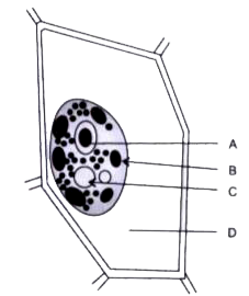
View SolutionGiven below is a diagram of the cell as seen under the microscope after having been placed in a solution.

What is the technical term used for the state/condition of the cell shown above?
View Solution
