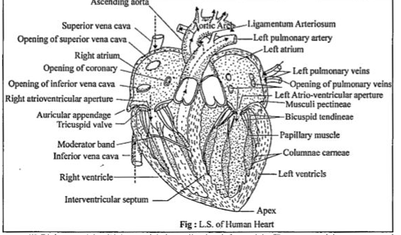
Describe the internal structure of human heart.
Text Solution
1) Right atrium : It is a thin walled chamber. It lies to the right and anterior to the left atrium. It is larger than the left atrium. On the lower margin, a muscular structure called auricular appendage hangs as a flap. Internally, atrium has a smooth walled venous part called sinus venarum. Right atrium recieves three large veins and number of small veins. These are superior vena cava, inferior vena cava, coronary sinus and small coronary veins. The superior vena cava opens in the right atrium. It brings the deoxygenated blood from head, neck and upper limbs. Inferior vena cava is larger than the superior vena cava. It brings deoxygenated blood from middle and lower parts of the body. It opens in the right atrium in lower region. Coronary sinus opens in the middle part of right atrium. It brings deoxygenated blood from walls of the heart. The opening of coronary sinus has valvular fold called valve of Thebesius. Some coronary veins open directly into right atrium by separate apertures.
ii) Left atrium : It is a cubical chamber. It lies behind the right atrium. It is separated from right atrium by an oblique septa. Left atrium also bears a muscular flap over its lower region. This is called auricular appendage. Internally, it also bears a smooth wall in the venous part called, sinus venarum. Left atrium recieves four pulmonary veins, two on each side. The left pair of pulmonary veins opens by a common aperture while right two pulmonary veins have different openings. These veins bring oxygenated blood from two lungs. A few very small coronary veins also opens here. Valves are absent on apertures of some veins but blood does not flow back into veins. It is due to presence of internal valves and presence of circular muscles near the apertures.

iii) Right ventricle: Right ventricle is smaller than left ventricle. The two ventricles are separated by an interventricular septum. Ventricular wall is thick and ridged internally. A muscular band connects the septum with the wall of ventricle. It is called moderator band. The surface of the ventricle possesses a number of irregular muscular ridges known as trabeculae carnae. There are also large papillary muscles. Papillary muscles are continued at the other end with collagenous cords called chordae tendineae.
Right atrium opens into right ventricle by an aperture called right atrioventricular or AV aperture. Right AV aperture is triangular in shape. It is guarded by valve called tricuspid valve. The valve possesses three membranous flaps. These flaps are connected to papillary muscles by inelastic bands called chordae tendineae. From the upper left corner of right ventricle develops pulmonary aorta. The opening is guarded by pulmonary valve. The valve enables the blood flow from right ventricle into pulmonary aorta.
iv) Left ventricle: It is larger than right ventricle. Its wall is thicker than right ventricle. The left atrium opens into left ventricle by a left atrioventricular (AV) aperture. The aperture bears bicuspid valve. The two flaps are attached to the rim of left AV aperture. From the right region of left ventricle, there arises systematic aorta.
|
Similar Questions
Describe the internal structure of human ear.
View SolutionDraw the internal structure of human heart.
View SolutionDescribe internal structure of a human heart.
View SolutionDescribe internal structure of a human heart.
View SolutionDescribe the internal structure of kidney.
View SolutionDraw a diagram to show the internal structure of human heart. Label only one heart chamber, any two heart valves and any three other structures.
View SolutionDescribe the structure of human heart.
View SolutionDescribe the structure of human heart.
View SolutionDraw a well labelled diagram of internal structure of human heart.
View Solution(a)Draw a labelled diagram of internal structure of human heart.
(b) What are chordae tendinae? Give their function.
View Solution
