Explain briefly repiratory system of cockroach.
Text Solution
Respiratory system. It is the system which is concerned with the exchange of gases and oxidation of food due to which CO2,H2O and energy are liberated.
C6H12O6+6O2−+6CO2+6H2O+Energy.
Unique features of Respiratory system :
1. It is the most efficient type because oxygen comes indirect contact with tissue cells due to tracheoles.
2. Blood does not take part in respiration.
3. Tissue fluid plays an important role in exchange of gases.
Organs of respiration. In cockroach the respiratory organs are :
1. Stigmata 2. Air tubes or tracheae.
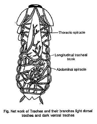
1. Stigmata : Tracheae communicate with outside by ten pairs of stigmata or spiracles. The stigmata lie along the sides of body in the soft pleura between terga and stema. There are two pairs of large thoracic stigmata-one pair lying between protorax and mesothorax and other lying between mesothorax and metathorax. The remaining eight pairs are small abdominal stigmata. There lie a pair each on the sides offirst eight abdominal segments near their anterior borders. The stigmata of first thoracic and first abdominal segment remain always opened whereas the remaining stigmata remain closed during expiration. The stigmata bear bristles which prevent the dust particles from entering into the body.
2. Tracheae. Each stigmata·opens into a small tracheal chamber or atrium from which arise trachael trunks. Atrium has bristly plates which check dust particles. From these trunks arise large tracheae which branch and rebranch forming a network. Their ultimate branches are called tracheoles which penetrate all the parts of body and end in tissue cells.
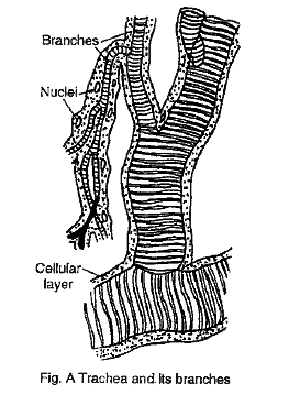
Each trachea is ectodermal in origin. It is formed by invagination of the integument. Its wall is composed of single layer of squamous epithelium surrounded by a delicate protoplasmic basement membrane. The epithelium is lined internally by a cuticular thickening arranged in a spiral manner. This thickening supports the trachea and prevents

it from collapsing. It is also shed during mouling. Thetracheoles have no cuticularthickneings. Their cavities are intracellular. They penetrate the tissue cells. They are partly filled with tissue fluid.
Tracheal end cell. Each tracheole ends within a cell called Tracheal end cell. From these cells arise very fine branches. These branches end blindly.

Inspiration. When the tergo-sternai muscles relax the abdomen is enlarged. Thus air from outside goes into the tracheae through stigmata. From tracheae air goes to tracheoles. Oxygen of air is dissolved into the tissue fluid and is then diffused into tissue cells. The intake of oxygen is called inspiration. Oxidation takes place in the tissue cells. Energy is produced, water and carbon dioxide are formed as end products.
Expiration. When tergosternal muS\.!les contract the abdomen is shortened. The tracheae are compressed therefore carbon dioxide is passed out through stigmata. Which is called expiration. The alternate and rhythmic contractions and expansions of the abdomen are called respiratory movements. Most of the carbon dioxide diffuses into the surrounding blood and is ultimately diffused out through through the cuticle which is impervious to water. The blood takes no part in respiration as the air reaches the tissues directly through the tracheae.
Nervous system. It is the system which is concerned to control and to co-ordinate all the body activities. The nervous system of cockroach consists of three parts :
1. Central nervous system
2. Peripheral nervous system
3. Sympathetic nervous system.
1. Central nervous system. It is that system which lies on median longitudinal axis of body. It consists of the following parts :
(a) Supra-oesophageal ganglion or cerebral ganglia or Brain. It lies dorsally in head region above the oesophagus. It is a bilobed mass. It is formed by fusion of three pairs of ganglia.
(b) Sub-oesophageal ganglion. It also lies in headregion ventral to the oesophagus. It is also formed by the fusion of three pairs of ganglia.
(c). Circum-oesophageal Connectives. These are a pair of flattened bands which arise from the sides of supraoesophageal ganglion. They run backwards and downwards. After encircling the oesophagus they unite laterally with the suboesophageal ganglion.
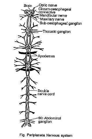
(d) A ganglionated double ventral nerve cord. From the sub-oesophageal ganglion arises posteriorly a double ventral nerve cord. The two parts of the cord remain separate. The cord runs backwards along the midventral line in the thorax and abdomen. It bears nine ganglia i.e. three thoracic and six abdominal. There is one ganglion in each thoracic segment. The first four abdminal ganglia lie in first four abdominal segments, one in each. The fifth abdominal ganglia lies in sixth segment. The sixth abdominal ganglion lies in seventh abdominal segment. All the thoracic ganglia and last abdominal ganglion are very large.
Peripheral nervous system :
Nerves arising from the central nervous system, constitute peripheral nervous system.
Nerves from brain. Following three pairs of nerves arise from the brain :
(i) Optic nerves. They go to the eyes.
(ii) Antennary nerves go to anthennae.
(iii) Labro frontal nerves go to labrum and frontal ganglion.
Nerves from sub-oesophageal ganglion :
The following four pair of nerves arise from the sub-oesophageal ganglion :

(i) Mandibular nerves go to mandibles.
(ii) Maxillary nerves go to maxillae.
(iii) Labial nerves go to labium.
(iv) Cervical nerves go to neck.
Several paired nerves arise from each thoracic ganglion. A pairofnerves arising from the ganglion of metathoracic segment runs backwards and supply the various parts of the first abdominal segment. Similarly each first five abdominal ganglia, send a pair of nerves to the 2nd, 3rd, 4th, 5th and 6th abdominal segments respectively. The 6th abdominal ganglion suppiies Several nerves to the. remaining 7 to 10 abdominal segments.
3. Sympathetic nervous system. It comprises the following ganglia :
(i) A small frontal ganglion. It lies in front of the brain. It is connected to brain by frontal connectives.
(ii) A pair of oesophageal ganglia. They lie behind the brain.
(iii) A large visceral ganglioii. It lies above the crop.
All the sympathetic ganglia are connecied to brain by connectives. A median unpaired recurrent nerve arises from the frontal ganglion and goes to the visceral ganglion or ingluvial ganglion.
Several nerves arising from these ganglia innervate the muscles, spicacles and alimentary canal.
|
Similar Questions
Knowledge Check
The circulatory system of cockroach
Ais present on dorsal side and has 3 chambers of heart in abdominal regionBis present on ventral side and has 10 abdominal and 3 thoracic chambers of heartCis present on ventral side and has 13 chambers of heart in abdominal regionDis present on dorsal side and has 10 abdominal and 3 thoracic chambers of heartCirculatory system of Cockroach is present
AOn dorsal side with 13 unit heart in abdomenBOn dorsal side with 10 abdominal and 3 thoracic units of heartCOn ventral side with 13 unit heart in abdomenDOn ventral side with 10 abdominal and 3 thoracic units of heartThe nervous system in cockroach -
AIs spread throughout the bodyBIs represented by segmentally arranged ganglia and ventral nerve cordCBoth a and b are correctDConsists of supra-oesopharyngeal ganglia in head and dorsal nerve cord
What is metamorphosis? Explain briefly metamorphosis in cockroach.
View SolutionMuschroom gland a part of-
(1) Digestive system of cockroach
(2) Respiratory system of cockroach
(4) Male resproductive system of earthworm
(4) Male resproductive system of cockroach
View SolutionExplain briefly, micturition and disorders of the excretory system.
View SolutionExplain briefly, micturition and disorders of the excretory system.
View SolutionExplain the nervous system of earthworm briefly.
View Solution
