Explain the parts of a flower, with a neat labelled diagram.Solution in Tamil
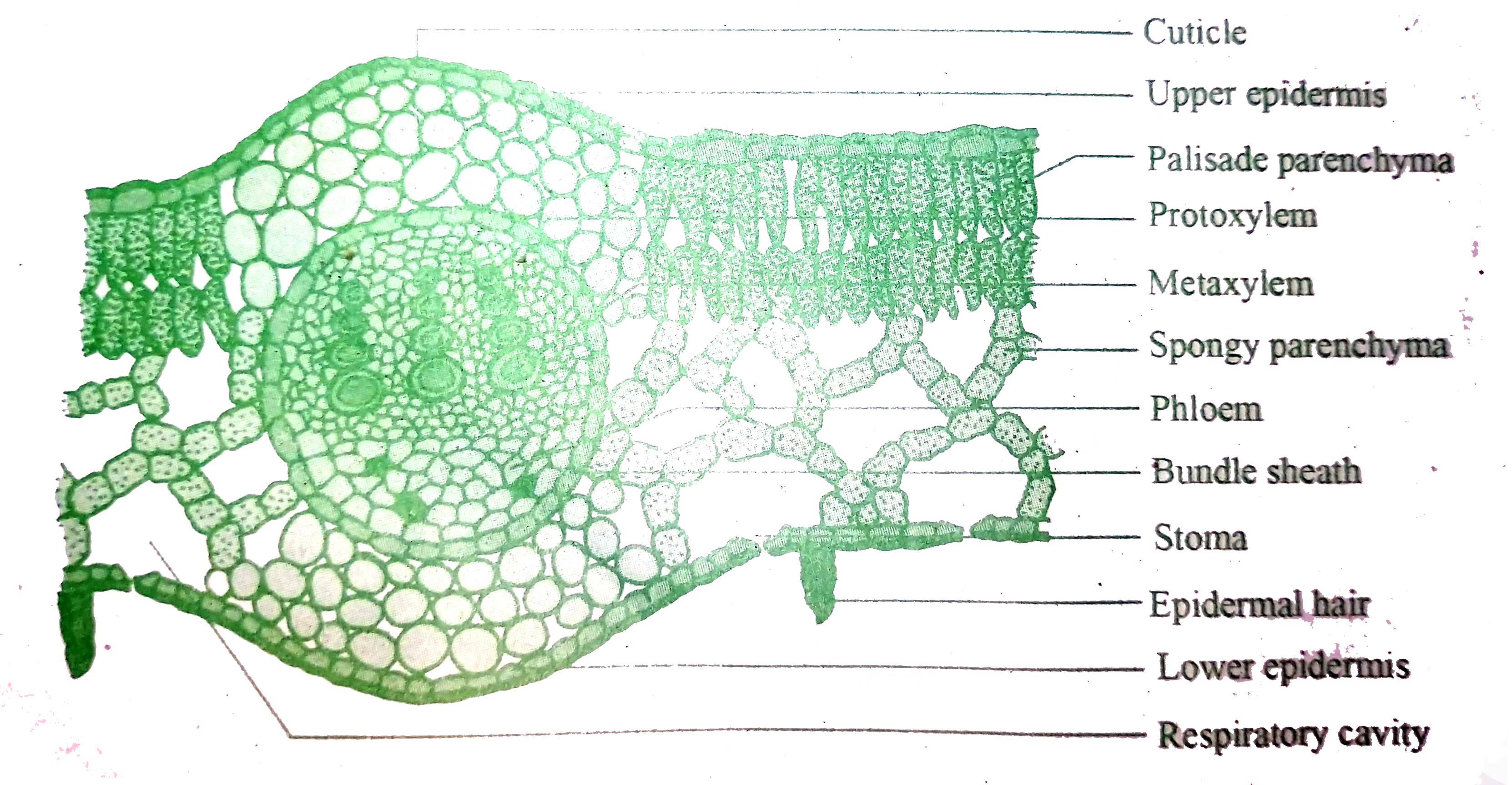
Anatomy of a Dicot Leaf -sunflower Leaf:Internal structure of dicotyledonous leaves reveal epidermis,mesophyll and vascular tissues.
Epidermis:This leaf is generally dorsiventral.It has upper and lower epidermis.The epidermis is usually made up of a single layer of cells that are closely packed.The cuticle on the upper epidermis is thicker than that of lower epidermis.The minute opening found on the epidermis are called stomata.Stomata are more in number on the lower epidermis than on the upper epidermis.A stomata is surrounded by a pair of bean shaped cells are called guard cells.Each stoma internally opens into an air chamber.These guard cells contain chlotroplasts .The main function of epidermis is to give protection to the inner tissue called mesophyll.The cuticle helps to check transpiration.Stomata are used for transpiration and gas exchange.
Mesophyll:The entire tissue between the upper and lower epidermis is called mesophyll (GK meso=in the middle,phyllome=leaf).There are two regions in the mesophyll. They are palisade parenchyma and spongy parenchyma.Paliside parenchyma cells are seen beneath the upper epidermis. It consists of vertically elongated cylindrical cells in one or more layers. These are compactly arranged and are generally without intercellular spaces. Palisade parenchyma cells contain more chloroplasts than the spongy parenchyma cells.The function of palisade parenchyma is photosynthesis.Spongy parenchyma lies below the palsied parenchyma. Spongy cells are irregularly shaped.These cells are very loosly arranged with numerous airspaces. As compared to palisade cells,the spongy cells contain number of chloroplasts.Spongy cells facilitate the exchange af gases with the help af air spaces.The air space that is found next to the stomata is called respiratory cavity or substomatal cavity.
Vascular tissue: Vascular tissue are present in the veins of leaf. Vascular bundles are conjoint collateral and closed. Xylem is present towards the upper epidermis,while the phloem towards the lower epidermis. Vascular bundles are surrounded by a compact layer by a parenchymatous cells called bundle sheath or border parenchyma.
Xylem consists of metaxylem and protoxylem elements.Protoxylem is present towards the upper epidermis,while the phloem consists of sieve tubes,companion cells and phloem parenchyma. Phloem fibres are absent.Xylem sonsists of vessels and xylem parenchyma.Tracheids and xylem fibres are absent.

Topper's Solved these Questions
- TISSUE AND TISSUE SYSTEMBOOK - PREMIERS PUBLISHERSCHAPTER - TISSUE AND TISSUE SYSTEMEXERCISE - OTHER IMPORTANT QUESTIONS & ANSWERS (CHOOSE THE CORRECT ANSWER)
40 Videos
- THE LIVING WORLD BOOK - PREMIERS PUBLISHERSCHAPTER - THE LIVING WORLD EXERCISE - OTHER IMPORTANT QUESTIONS & ANSWERS (Answer the following questions.)
52 Videos
- TISSUE LEVEL OF ORGANISATIONBOOK - PREMIERS PUBLISHERSCHAPTER - TISSUE LEVEL OF ORGANISATIONEXERCISE - SOLUTION TO TEXTUAL QUESTIONS
6 Videos
Similar Questions
Choose the correct answer.(1 mark)
Who is the father of plant anatomy?
View SolutionDefine ecosystem.
View SolutionStraified epithelia are built for the protection or to resist abration. How are the simple epithelia better at ?
View SolutionWith a neat labelled diagram, explain how a rainbow is formed.
View SolutionDraw the ultrastructure of mitochondria and label the parts.
View SolutionExplain myopia or short sighteness with a neat labelled diagram
View Solutionwith a neat labelled diagram, explain phloem tissue.
View SolutionExplain the structure of mitochondria with a neat labelled diagram.
View SolutionDraw a neat diagram to explain the exchange of gases at blood level and label the parts.
View SolutionWith a neat labelled diagram, explain phloem tissue.
View SolutionDraw a neat labeled diagram of T-S of a mature anther and explain the parts.
View SolutionExplain the parts of a flower, with a neat labelled diagram.
View SolutionWith a neat labelled diagram, explain the functions of the main parts of the human eye.
View SolutionDraw a neat labelled diagram of the human eye and explain the working of each part of it.
View SolutionExplain the components of a biogas plant with a neat labeled diagram.
View SolutionDraw a neat labelled diagram of stamen of typical flower.
View Solution
