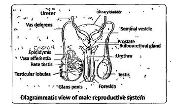
Describe the male reproductive system in Human.Solution in Telugu
The Male Reproductive System: The male reproductive system (male genital system consists of a number of sex organs that are a part of the human reproductive process. The sex organs which are located in the pelvic region include a pair oftestes (sing: testis) along with accessory ducts, glands and the external genitalia.
Testes : The testes (testicles) are a pair of oval pinkish male primary sex organs suspended outside the abdominal cavity with in a pouch called scrotum. The scrotum helps in maintaining the low temperature of the testes ( lower than the normal internal body temperature) necessary for spermatogenesis. The cavity of the scrotal sac is connected to the abdominal cavity through the inguinal canal. Testis is held in position in the scrotum by the gubernaculum, a fibrous cord that connects the testis with the bottom of the scrotum and a-spermatic cord, formed by the vas deferens, nerves, blood , vessels and other tissues that run from the abdomen down to each testicle, through the in guinalcanal . Each testis is enclosed in a fibrous envelope, the tunica albuginea, which extends inward to form septa. that partition the testis into lobules. There are about 250 testicular, lobules in each testis. Each lobule contains 1 to 3 highly coiled seminiferous tubules. A pouch of serous membrane (peritoneal layer) called tunica vaginal is covers the testis:

Each seminiferous tubule is lined by the germinal epithelium which consists of undifferentiated. male germ cells called spermatogonial mother cells and it also bears, nourishing cells called Sertoli cells. The spermatogonia produce the primary spermatocytes which undergo meiotic division, finally leading to the formation of spermatozoa or sperms (spermatogenesis). Sertoli cells provide nutrition to the spermatozoa and also produce a hormone called inhibin, which inhibits the secretion of FSH. The regions outside the seminiferous tubules, called interstitial spaces, contain interstitial cells of Leydig or Leydig cells. Leydig cells produce androgens, the most important of which is testosterone. Testosterone controls the development of secondary sexual characters and spermatogenesis. Other immunologically competent cells are also present. The seminiferous tubules open into the vasa efferentia through the rete testis (a network of tubules in of the testis carrying spermatozoa from the seminiferous tubules to the vasa efferentia).
Epididymnis : The vasa efferentia leave the testis and open into a narrow, tightly coiled tube called epididymis located along the posterior surface of each testis. The epididymis provides a storage space for the sperms and gives the sperms time to mature. It is differentiated into three regions-caput epididymis, corpus epididymis and cauda epididymis. The caput epididymis receives spermatozoa via the vasa efferentia of the mediastinum testis (a mass of connective tissue at the back of the testis that encloses the rate testis).
Vasa deferentia : The vas deferens or ductus deferens is a long, narrow, muscular tube. The mucosa of the ductus deferens consists of pseudostratified columnar epithelium and lamina propria (areolar connective tissue). It starts from the tail of the epididymis, passes through the inguinal canal into the abdomen and-loops over the urinary bladder. It receives a duct from the seminal vesicle. The vas deferens an a the duct of the seminal vesicle unite to form a short ejaculatory duct / ductus ejaculatorius. The two ejaculatory ducts, carrying spermatozoa and the fluid secreted by the seminal vesicles, converge in the centre of the prostate and open into the urethra, which transports the sperms to outside.
Urethra: in males, the urethra is the shared terminal duct of the reproductive and urinary systems. The urethra originates from the urinary bladder and extends through the penis to its external opening called urethral meatus. The urethra provides an exit for urine as well as semen during ejaculation me
Penis: The penis and the scrotum constitute the male external genitalia. The penis serves as urinal duct and also intromittent organ that transfers spermatozoa to the vagina of a female. The human penis is made up of three columns of tissue, two upper corpora cavernosa on the dorsal aspect and one corpus spongiosum on the venttal side. Skin anda subcutaneous layer enclose all three columns, which consist of special tissue that helps. in erection of the penis to facilitate insemination. The enlarged and bulbous end of penis called glans penis is covered by a loose fold of skin (foreskin) called prepuce. The urethra traverses the corpus spongiosum, and its opening, lies at the tip of the glans penis (urethral meatus).
Male accessory genital glands.:- The male accessory glands include paired seminal vesicles,, a prostate and bulbourethral glands.
Seminal vesicles : The seminal vesicles are a pair of simple tubular glands present postero- inferior to the urinary bladder in the pelvis. Each seminal vesicle opens into the corresponding vas deferens, as the vas deferens enters the prostate gland: The secretion of the seminal vesicles constitutes about 60 percent of the volume of seminal fluid. It is an alkaline, viscous fluid that contains fructose, proteins, citric acid, inorganic phosphorus, potassium, and prostaglandins. Once this fluid joins the sperm in the ejaculatory duct, fructose acts as the main energy source for the sperm outside the body. Prostaglandins are believed to aid -fertilization by causing the mucous lining of the cervix to be more receptive to sperm as well as by aiding the movement of the sperm towards the ovum with peristaltic contractions of the uterus and fallopian tubes.
Prostate gland: Prostate gland is located directly beneath the urinary bladder. The gland surrounds the prostatic urethra, and sends its secretions through several prostatic ducts. In man, the prostate contributes-15 - 30 percent of the semen. The fluid from the prostatic is clear and slightly acidic. The prostatic secretion .activates. the spermatozoa and provides nutrition.
Bulbourethral Glands, Bulbourethral glands, also called Cowper.s glands, are located beneath the prostate gland at the beginning of the internal portion of the penis. They add an alkaline fluid to semen during the process of ejaculation. The fluid secreted by these glands lubricates the urethra. It is also thought to function as a .flushing agent. that washes out the acidic urinary residues that renain in the urethra, before the semer is ejaculated.
Topper's Solved these Questions
Similar Questions
Define and explain water potential.
06:09View SolutionDescribe the structure of the heart of a man. Draw a neat labelled diagram of it.
View SolutionGive an account of glycolysis, Where does it occur ? What are the end products? Trace the fate of these products in both aerobic, and anaerobic respiration.
09:04View SolutionDraw a labeled diagram of male reproductive system.
View SolutionWith the help of a neat and labelled diagram, describe the human male reproductive system.
View SolutionDescribe the human male reproductive system with the help of a neat labelled diagram.
View SolutionDescribe the male reproductive system.
07:27View SolutionDescribe the male reproductive system.
05:26View Solutionमनुष्य में नर जनन तन्त्र का वर्णन कीजिए।
03:34View SolutionDescribe the structure of male reproductive system in human beings.
05:10View Solutionమానవ పురుష ప్రత్యుత్పత్తి వ్యవస్థను వర్ణించండి.
08:06View SolutionDescribe the hormonal control of reproductive system in human male.
01:40View SolutionDescribe the three parts of male reproductive system in human
10:06View SolutionDescribe the human male reproductive system with well labelled diagram.
08:43View SolutionDescribe with diagram : Human - male reproductive system
02:30View SolutionDescribe the human male reproductive system with the help of a neat labelled diagram.
01:01:12View Solution

