The inner most elongated cells of ommatidium are
rhabdomere
cone cells
photoreceptor cells
cornea
The correct Answer is:C
To solve the question regarding the innermost elongated cells of the ommatidium, we can follow these steps:
1. Understand the Structure of Ommatidia:
- Ommatidia are the basic structural units of the compound eye found in insects, such as cockroaches. Each ommatidium is composed of several types of cells that play specific roles in vision.
2. Identify the Types of Cells in Ommatidia:
- The main types of cells found in an ommatidium include:
- Corneogen cells: These are responsible for secreting the cornea, which is the outermost layer of the eye.
- Raptomeres: These are elongated cells and are the innermost cells of the ommatidium.
- Cone cells: These cells are involved in light vision but are not the innermost cells.
- Photoreceptor cells: These cells contribute to the overall sight but are not classified as the innermost cells.
3. Determine the Function of Each Cell Type:
- Corneogen cells → Secrete the cornea (outermost layer).
- Raptomeres → Elongated and innermost cells.
- Cone cells → Help in vision under light conditions.
- Photoreceptor cells → Aid in overall sight.
4. Identify the Correct Answer:
- Since the question specifically asks for the innermost elongated cells of the ommatidium, the correct answer is raptomere.
Final Answer:
The innermost elongated cells of ommatidium are raptomere.
---
- Ommatidia are the basic structural units of the compound eye found in insects, such as cockroaches. Each ommatidium is composed of several types of cells that play specific roles in vision.
- The main types of cells found in an ommatidium include:
- Corneogen cells: These are responsible for secreting the cornea, which is the outermost layer of the eye.
- Raptomeres: These are elongated cells and are the innermost cells of the ommatidium.
- Cone cells: These cells are involved in light vision but are not the innermost cells.
- Photoreceptor cells: These cells contribute to the overall sight but are not classified as the innermost cells.
- Corneogen cells → Secrete the cornea (outermost layer).
- Raptomeres → Elongated and innermost cells.
- Cone cells → Help in vision under light conditions.
- Photoreceptor cells → Aid in overall sight.
- Since the question specifically asks for the innermost elongated cells of the ommatidium, the correct answer is raptomere.
The innermost elongated cells of ommatidium are raptomere.
Topper's Solved these Questions
- STRUCTURAL ORGANISATION IN ANIMALSBOOK - AAKASH SERIESCHAPTER - STRUCTURAL ORGANISATION IN ANIMALSEXERCISE - EXERCISE-I (COCKROACH) (REPRODUCTORY SYSTEM)
29 Videos
- STRUCTURAL ORGANISATION IN ANIMALSBOOK - AAKASH SERIESCHAPTER - STRUCTURAL ORGANISATION IN ANIMALSEXERCISE - EXERCISE-I (FROG)(EXTERNAL CHARACTERS)
13 Videos
- STRUCTURAL ORGANISATION IN ANIMALSBOOK - AAKASH SERIESCHAPTER - STRUCTURAL ORGANISATION IN ANIMALSEXERCISE - EXERCISE-I (COCKROACH) (NERVOUS SYSTEM)
14 Videos
- STRATEGIES FOR ENHENCEMENT IN FOOD PRODUCTIONBOOK - AAKASH SERIESCHAPTER - STRATEGIES FOR ENHENCEMENT IN FOOD PRODUCTIONEXERCISE - EXERCISE-III
68 Videos
- THE LIVING WORLD BOOK - AAKASH SERIESCHAPTER - THE LIVING WORLD EXERCISE - EXERCISE-III (Previous AIPMT/NEET Questions)
12 Videos
Similar Questions
Knowledge Check
What is formed by corneagen cells of an ommatidium?
ACone cellsBCrystalline coneCLensDNone of theseInternode elongation is due to
AgibberellinsBAuxinsCcytokininDabscisic acid
Give the technical terms for the
Elongated cells which are thickened at the corners.
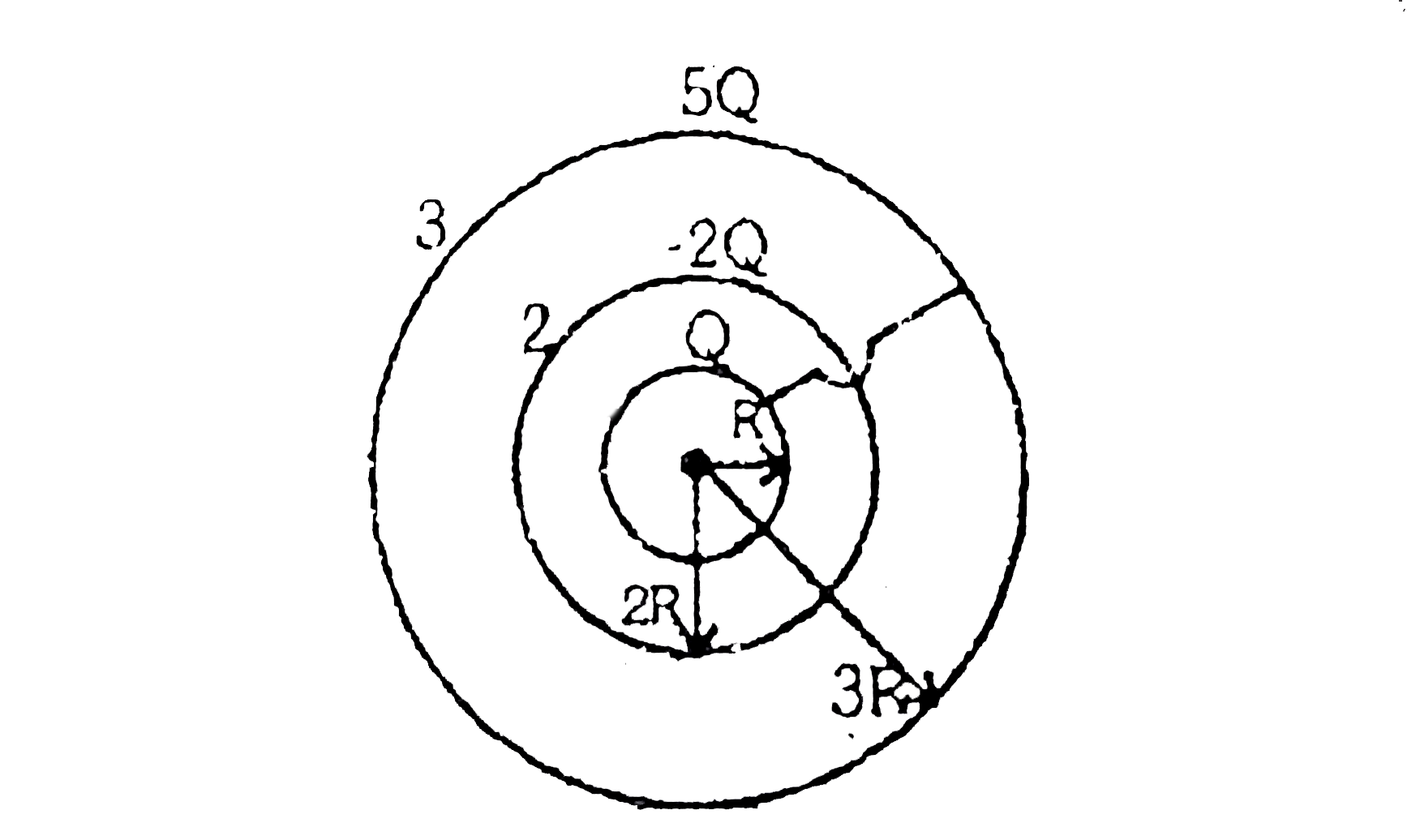
View SolutionFind charge on each spherical shell after joining the inner most shell outer most shell by a conducting wire. Also find charges on each surface.

View Solution[A]: The inner most distinict layer of the cortex is called endodermis.
(R]: The cells of endodermis are nonliving and bear casparian strips
View SolutionTracheids are elongated cells with thick and (A)_____ walls and (B)_____ ends.
View SolutionThe inner wall of each guard cell, towards the pore or stomatal aperture, is
View SolutionThe inner wall of each guard cell, towards the pore or stomatal aperture, is
View Solution
