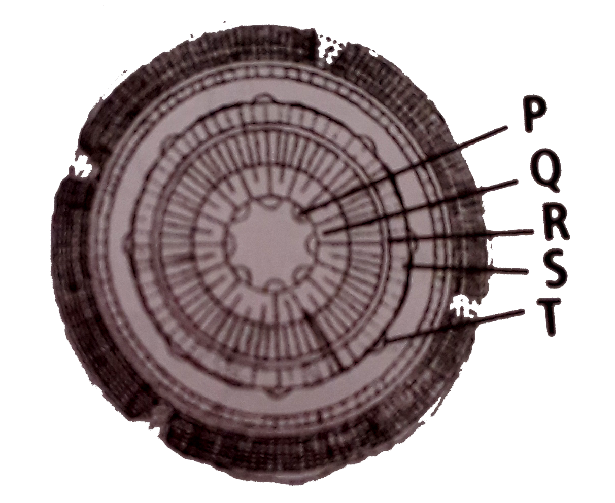
Describe the T.S. of Dicot Stem.
Text Solution
Internal structure of young dicot stem- Ex Helianthus annuus
Transverse section of a dicot stem shows three distinct zones -epidermis, cortex and stele.
1. Epidermis:
1. The outermost layer in young dicot stem is called epidermis. It is one cell in thickness.
2. Cells are tubular or rectangular.They are arranged compactly without interacellular spaces.
3. Outer surface of epidermis is covered by a waxy subtance called cutin. This layer is caled cuticle.
4. Minute pores found in the epidermis are caled stomata.
Multicellular hairs developing onthe epidermis are called trichomes.
6.Epidermis gives protection to the inner tissues.
7. Stomata facilitates exchange of gase and promotes transpiration.

8. Cuticle and trichomes check transpiration. They also protect the stem from high temperature.
9.Trichomes also help in preventing the enrty of pathogenic micro-organisms.
II. Cartex: It is exrtrastelar ground tissue. It shows three subzones -hypodermis, general cortex and endodermis.
A. Hypodermis:
1. The layer present below the epidermis is called hypodermis.
2.It consist of 3-6 layers of Collenchyma.
3. Cells are arranged compactly without intercellular spaces.
They shows excessively thickened corners.
4.Hypodermis helps in providing tensile strength to the stem.
5. Hypodermis also helps in production of food materials by haivng chloroplasts.
B.Genral Cortex:
1.It is found below the hypodermis.
2. It consists of 5-10 rows of parenchyma.
3.Cells are isodiametric or oval or spherical.
4.Resin or latex ducts may be present in it.
5. Outer layers of cells have chloroplasts and perform assimilation of food materials.
6. Inner layers are concerned with storage of food.
C.Endodermis:
1. It is the innermost layer of cortex.
2.It is one layer barrel shaped, compactly arragned cells.
3. Radial and transverse walls show casparian bands.
4. Endodermal cells store starch grains. So it is known as starch sheath.
III. Stele: Central conducting cylinder is called stele. It occupies major portion of stem. it shows four parts.
a. Pericycle:
1. It is the outermost layer of stele.
2. It lies betwen endodermis and vascular bundles.
3.It has alterate patches of sclerenchyma and parenchyma.
B. Vascular Bundles:
1. Each vascular bundle of vascular bundles are arranged in the shaped. 2. Limited number of vascular bundles are arranged in the shape of a circular ring. Such arrangement is called Eustele.
3. In each vascular bundle the phloem is present outside and xylem towards inside on the same radius. So vascular bundle is conjoint and collateral.
4.Meristematic tissue is present in betwen xylem and phloem. It is called fascicular cambium. Vascular bundle with cambium is called open type.
5.Xylem is endarch (protoxylem towards centre)
6. Xylem has vessels and xylem parentchyma. Tracheids and fibres are also present Xylem conducts water and salts.
7. Phloem has sieve tubes, companion cells, phloem parenchyma and fibres. It conducts food materials.
C. Medulla: Central part of stele is called medula. It is filled with parenchyma. It is well developed and extensive. It stores food materials.
D. Medullary rays: Medulla extends tothe periphery in between the vascular bundles forming medullary rays. Parenchymatous cells are living thin walled and elongate radially.
Medullary rays connect stele and cortex. They help in lateral conduction.
|
Updated on:21/07/2023
Topper's Solved these Questions
Similar Questions
Knowledge Check
- Question 1 - Select One
In dicot stem
AVascular bundles are conjoint, open and arranged in a ringBXylem is endarchCCollenchymatous hypodermisDAll - Question 2 - Select One
A.T.S. of dicot stem shows
Avascular bundles arranged in a ringBscattered vascular bundles.Cclosed vascular bundlesDradial vascular bundles.

