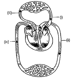
The given figure shows a section of the heart. Select the option that correctly matches the labels P, Q, R, S in the figure with the given list (i - iv) indicating direction of movement of blood.
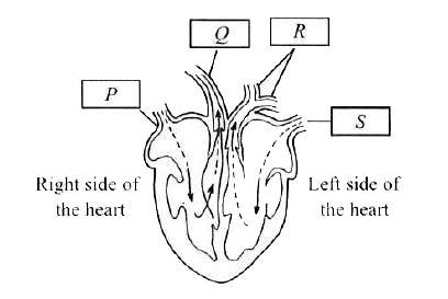
(i) To rest of the body
(ii) To lungs
(iii) From the lungs
(iv) From the rest of the body
P - (iv), Q - (i), R - (ii), S - (iii)
P - (iii), Q - (ii), R - (i), S - (iv)
P - (iv), Q - (ii), R - (i), S - (iii)
P - (iii), Q - (i), R - (ii), S - (iv)
The correct Answer is:C
Topper's Solved these Questions
- NSO MODEL TEST PAPER 03Book:SCIENCE OLYMPIAD FOUNDATION Chapter:NSO MODEL TEST PAPER 03Exercise:ACHIEVERS SECTION
Explore 3 Videos
- NSO MODEL TEST PAPER 02Book:SCIENCE OLYMPIAD FOUNDATION Chapter:NSO MODEL TEST PAPER 02Exercise:ACHIEVERS SECTION
Explore 4 Videos
- NSO QUESTION PAPER 2017- 18 SET - BBook:SCIENCE OLYMPIAD FOUNDATION Chapter:NSO QUESTION PAPER 2017- 18 SET - BExercise:ACHIEVERS SECTION
Explore 2 Videos
Similar Questions
Knowledge Check
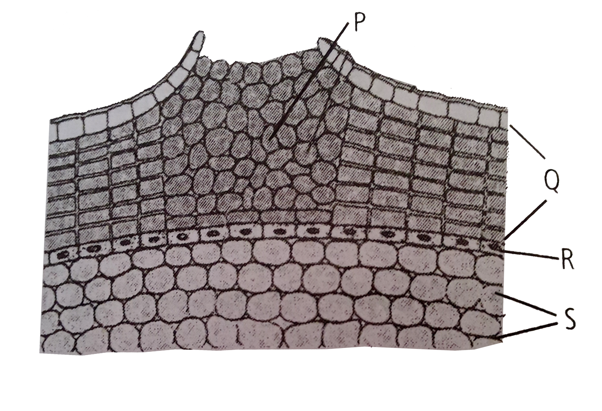
In the given transverse section of stem showing periderm, identify the parts labelled P, Q, R, S and select the correct option.APQRSComplementary cellsCorkPhellogenPhellodermBPQRSLenticelsCorkPhellodermPhellogenCPQRSLenticelsPhellodermPhellogenCorkDPQRSComplementary cellsPhellodermPhellogenCorkIdentify the joints labelled as P, Q, R and S in the given figures and select the correct option.
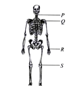 AGliding jointBall and socket jointHinge jointPivot jointSPQRBGliding jointBall and socket jointHinge jointPivot jointRQPSCGliding jointBall and socket jointHinge jointPivot jointRQSPDGliding jointBall and socket jointHinge jointPivot jointPSQR
The given figure shows a section of brain. Identify the parts labelled as A,B,C and D and match them with the names (i-vii) given below.
(i)Arachnoid membrane , (ii)Subdural space , (iii)Duramater , (iv)Bone , (v)White matter , (vi)Grey matter , (vii)Piamater
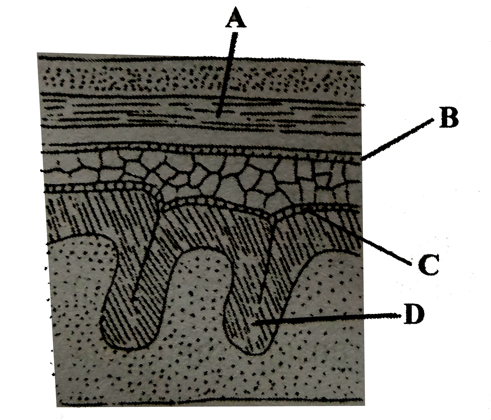 AABCD(iii)(ii)(vi)(v)BABCD(i)(ii)(iii)(vi)CABCD(iii)(i)(vii)(vi)DABCD(iv)(vii)(i)(ii)
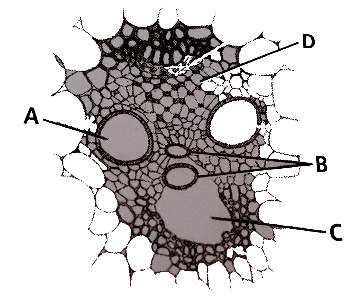
Refer to the given figure which represents a section of vascular bundles as seen in T.S. of a monocot stem and select the option that correctly labels A, B, C and D
View Solution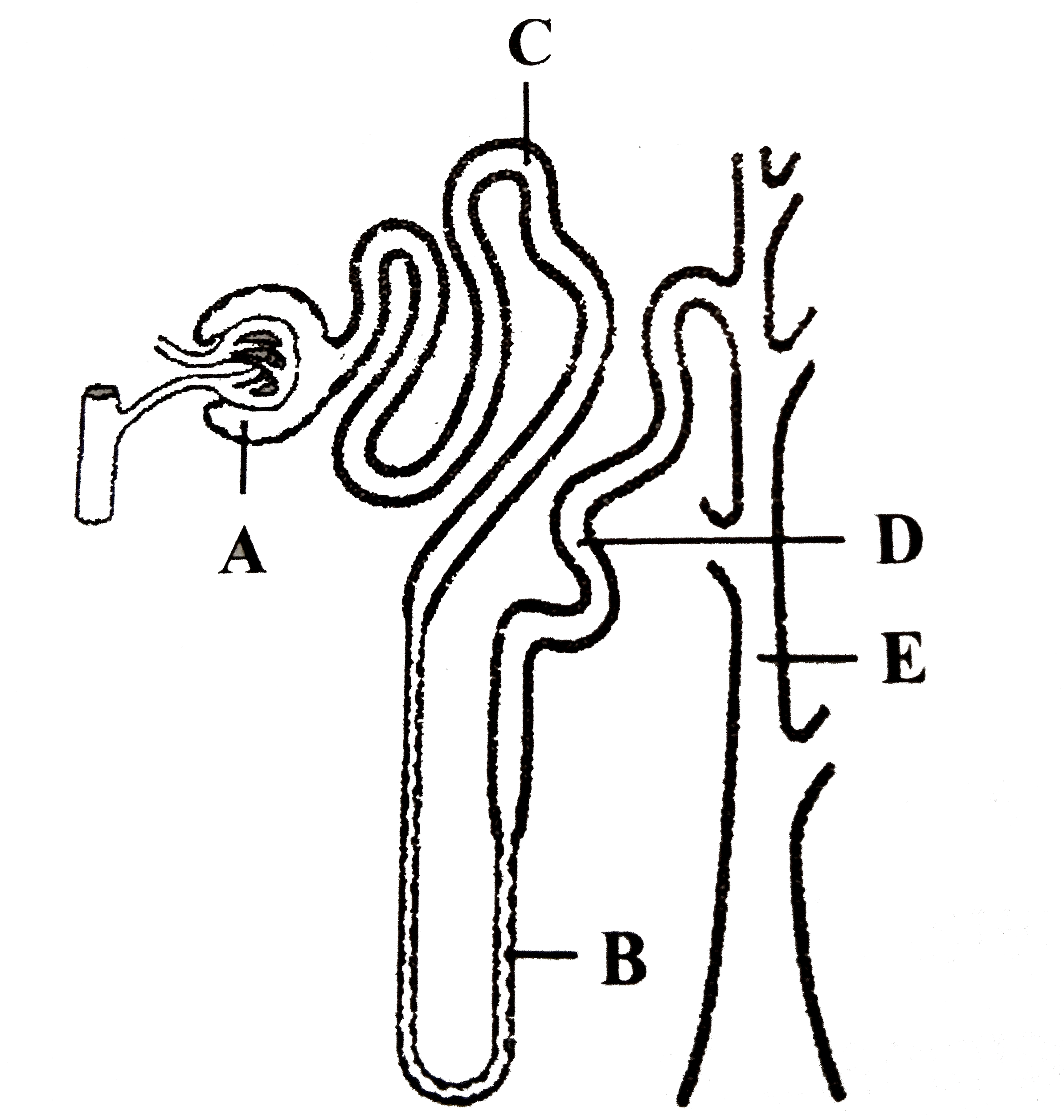
The given figure represents a single nephron from a mammalian kidney. Identify the labelled parts, match them with the functions (i-iv) and select the correct option.
(i). The site of ultrafiltration.
(ii). Particularly sensitive to ADH.
(iii). The main site for the reabsorption of glucose and amino acids.
(iv). Largely responsible for the maintenance of blood pH.
View SolutionMatch I with II and select the correct option from the given codes.

View SolutionThe given figure illustrates the changes in lung volume during the process of breathing.
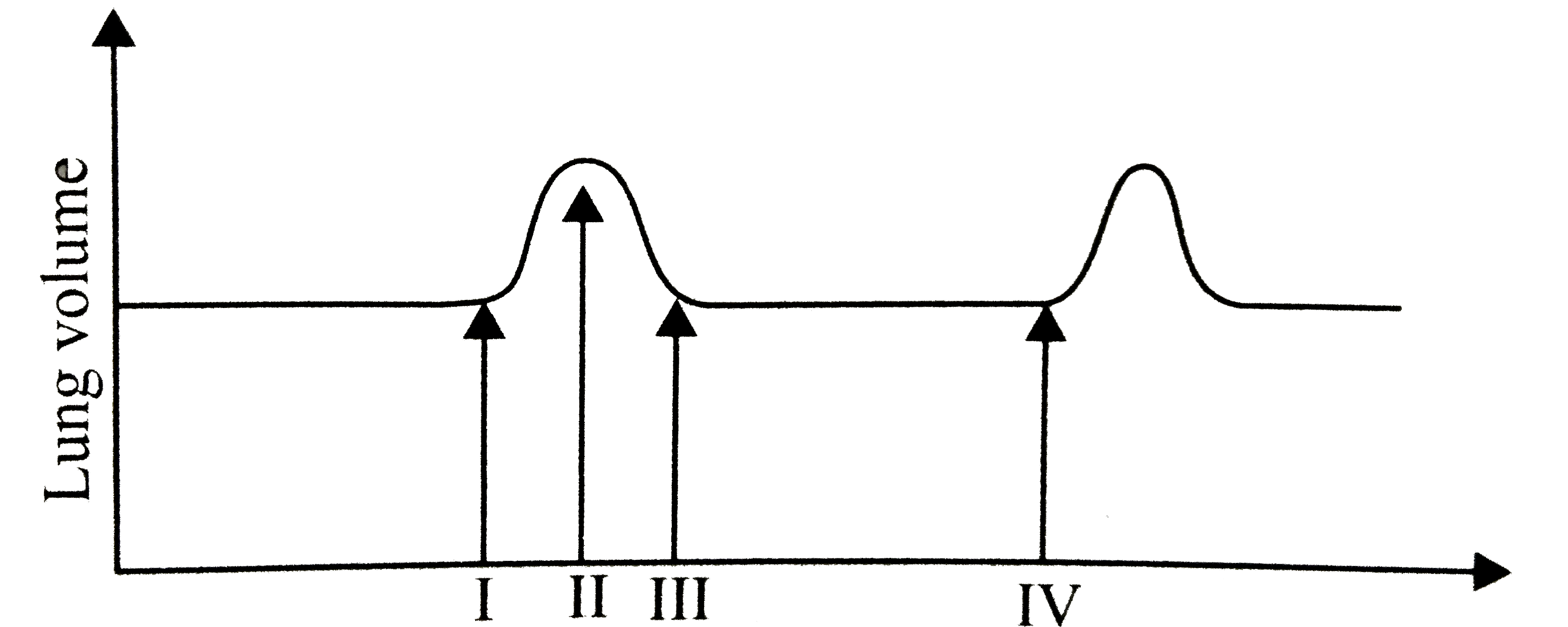
The changes from II to III indicates the
View SolutionThe figure given below shows a schematic plan of blood circulation in humans with labels (i) to (iv). Identify the correct label with its functions ?
View Solution

