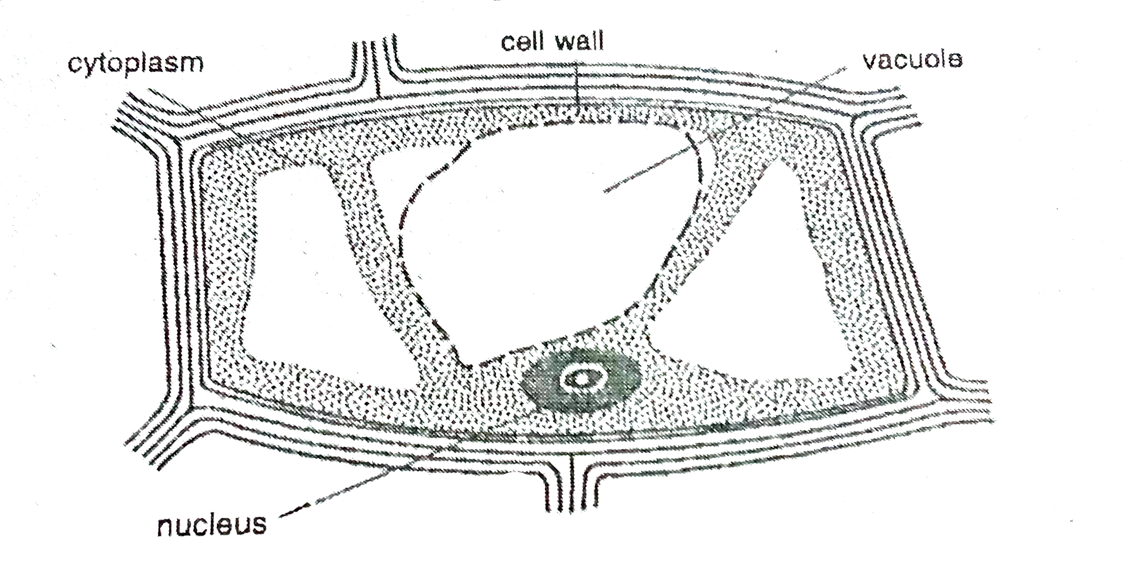
A student prepared a temporary mount of a leaf peel and observed some green dots.
(i) Name these green dots and name the pigment present in them.
(ii) State the function of this pigment.
(i) Green dots are chloroplast. It contains green pigment called chlorophyll.
(ii) Chlorophyll traps solar energy. This solar energy is required by the plant to carry out the chemical reactions involved in the preparation of food ( i.e., in photosynthesis ) .
Topper's Solved these Questions
- X BOARDSBOOK - X BOARDSCHAPTER - X BOARDSEXERCISE - SET-I SECTION-A
1 Videos
- X BOARDSBOOK - X BOARDSCHAPTER - X BOARDSEXERCISE - SET-I SECTION-B
1 Videos
- X BOARDSBOOK - X BOARDSCHAPTER - X BOARDSEXERCISE - SET III (SECTION -C)
1 Videos
- SAMPLE QUESTION PAPER BOOK - X BOARDSCHAPTER - SAMPLE QUESTION PAPER EXERCISE - Section-B
11 Videos
Similar Questions
Knowledge Check
While preparing a good temporary mount of leaf peel to observe stomata, care should be taken to avoid:
Aadding glycerine to the slideBusing water to wash the slideChaving air bubbles in the slideDstaining the peel with safraninTo prepare stained temporary mounts of (a) onion peel, and (b) human cheek cells and to record observations and draw their labelled diagram.


The structural or functional unit of life isAtissueBorganCorgan systemDcell
List the steps of preparation of temporary mount of a leaf peel to observe stomata.
View SolutionName the green pigment present in the leaves of a plant
View SolutionTo prepare a good temporary mount of the petunia leaf peel showing many stomata, from where does the student has to get the peel?
View SolutionTo prepare a good temporary mount of the petunia leaf peel showing many stomata, from where does the student has to get the peel?
View SolutionA student is observing the temporary mount of a leaf peel under a microscope. Draw labelled diagram of the structure (of stomata) as seen under the microscope.
View SolutionA student is observing the temporary mount of a leaf peel under a microscope. Draw
labelled diageram of the structure (of stomata) as seen under the microscope.
View Solution
