With the help of suitasble diagrams, explain in mitosis in detail.
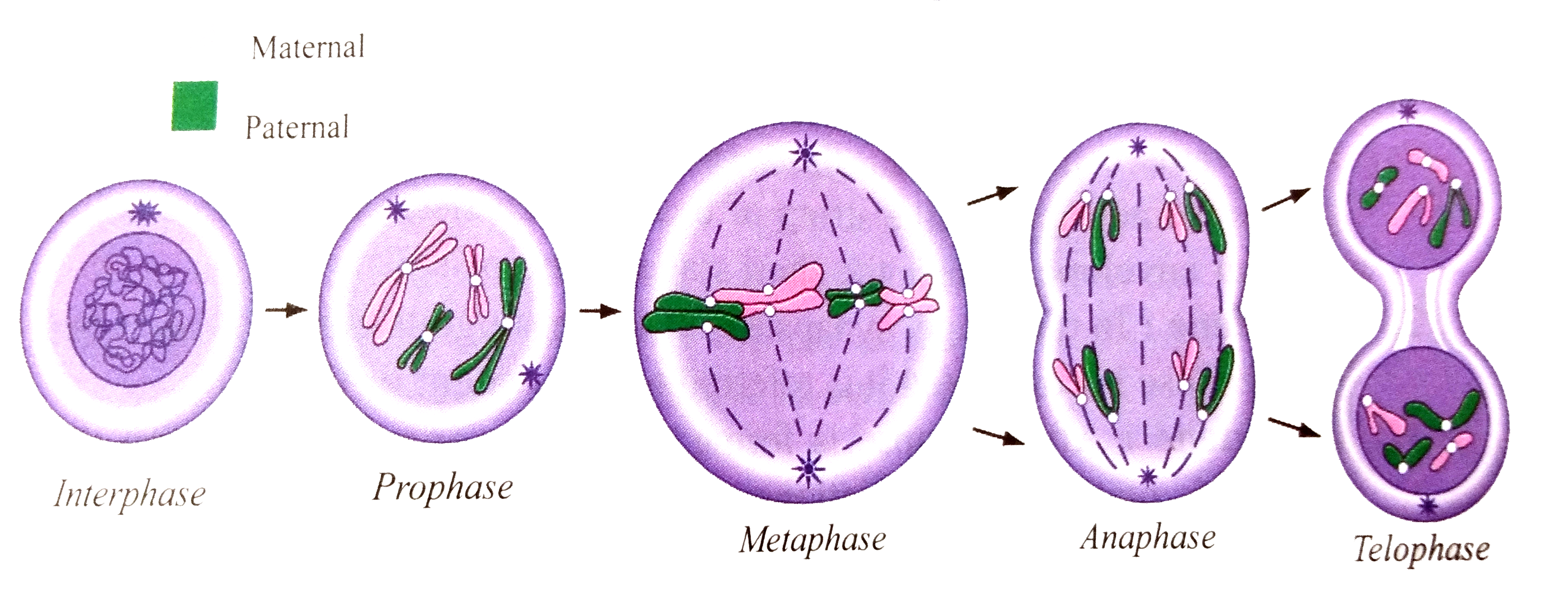
(1) There are two stages of mitosis. These are (a) Karyokinesis or nuclea rdivision and (b) Cytokinesis or cytoplasmic division. Karyokinesis takes places in further four phases, viz prophase, metaphase, anaphase and telophase.


a. Karyokinesis:
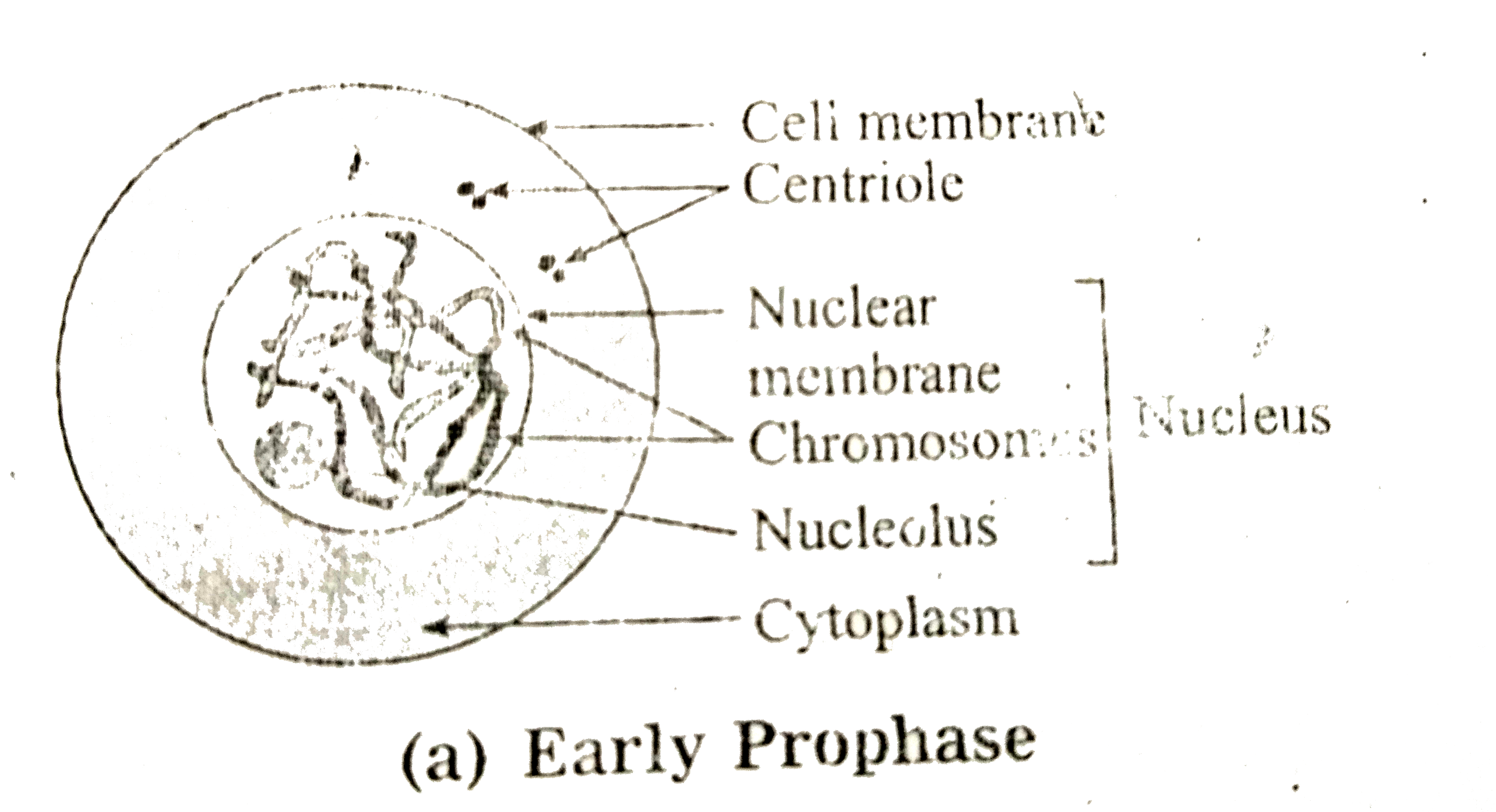
(i) Prophase: During prophase, condensation of chromosomes starts. The thin and thread like chromosomes start thickening. They are seen with their pair of sister chromatides. In animal cells the centroioles are seen to duplicate and move to opposite poles of the cell. uclear membrane and nucleolus disappear.
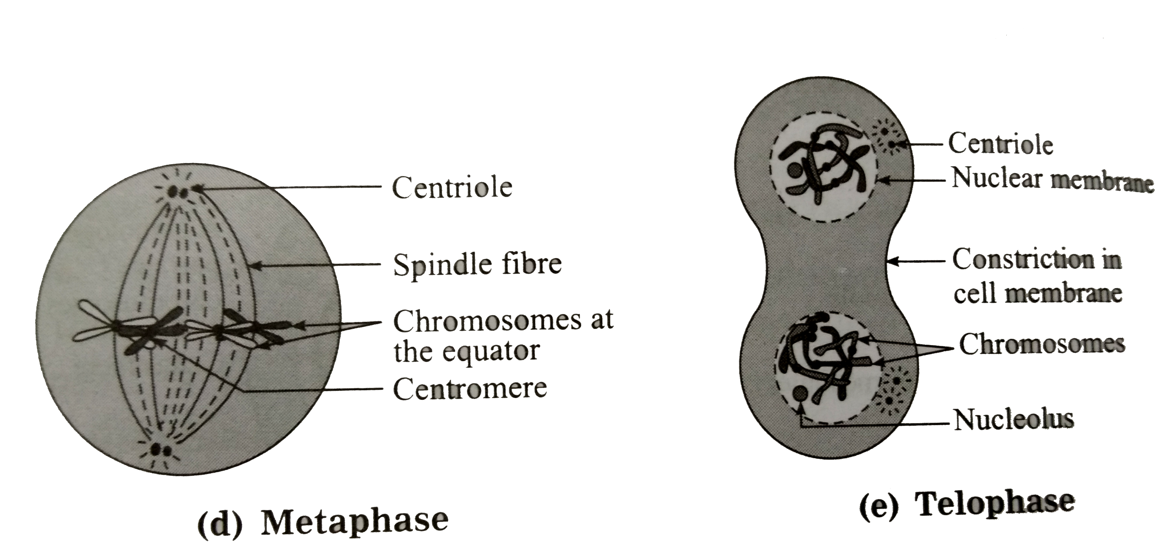
(ii) Metaphase: Chromosomes complete their condensation and each one is seen with its sister chromatids. The chromosomes are seen in equatorial plane of the cell. The spindle fibres are formed from polar region, where centrioles are present, and they attach themselves to the centromere of each chromosome. Nuclear membrane now disappears completely.
(iii) Anaphaese: The centromeres of the chromosomes now divide forming two daughter chromosomes. The spindle fibres pull apart the chromosomes from equatorial region to the opposite poles. Chromosomes mvoing to the poles appear like bunch of bananas. One set of chromosomes reach each pole by the end of the anaphase.
(iv) Telophase: Telophase is reverse of events that occurred in prophase. The thickened chromosomes decondense. They again assume the thin and thread like appearance. Nuclear membrane and nucleolus appear again. The spindle fibres are completely lost. The cell looks as if it has to nuclei in one cytoplasm.
b. Cytokinesis: In animal cells a notch develops in the middle of the cell. This notch goes ondeepening down and later the cytoplasm divides into two. In plants cells, cell plate formation takes place and then cytokinesis takes place.

a. Karyokinesis:
(i) Prophase: During prophase, condensation of chromosomes starts. The thin and thread like chromosomes start thickening. They are seen with their pair of sister chromatids. In animal cells the centrioles are seen to duplicate and move to opposite poles of the cell. Nuclear membrane and nculeolus disappear.
(ii) Metaphase: Chromosomes complete their condensation and each one is seen with its sister chromatids. The chromosomes are seen inequatorial plane of the cell. The spindle fibres are formed from polar region, where centrioles are present, and they attach themselves to the centromere of each chrosome. Nuclear memberane now disappears completely.
(ii) Anaphase: The centromeres of the chromosomes now divide forming two daghter chromosomes. The spindle fibres pull apart the chromosomes from equatorial region to the opposite poles. Chromosomes moving to the poles appear like bunch of bananas. One set of chromosomes reach each pole by the end of the anaphase.
(iv) Telophase: Telophase is reverse of events that occurred in prophase. The thickened chromosomes decondense. They again assume the thin and thread like appearance. Nuclear membrane and nucleolus appear again. The spindle fibres are completely lost. The cell looks as it has two nuclei in one cytoplasm.
b. Cytokinesis: In animal cells a notch develops in the middle of the cell. This notch goes on deepening down and later the cytoplasm divides into two. In plant cells, cell plate formation takes place and then cytokinesis takes place.
Topper's Solved these Questions
- SKETCHING, EXPLANATION, CORRECTION OF DIAGRAMSBOOK - NAVNEET PUBLICATION - MAHARASHTRA BOARDCHAPTER - SKETCHING, EXPLANATION, CORRECTION OF DIAGRAMSEXERCISE - SKETCH AND LABEL THE GIVEN DIAGRAMS:
3 Videos
- SKETCHING, EXPLANATION, CORRECTION OF DIAGRAMSBOOK - NAVNEET PUBLICATION - MAHARASHTRA BOARDCHAPTER - SKETCHING, EXPLANATION, CORRECTION OF DIAGRAMSEXERCISE - Observe the above figure and answer the questions below:
5 Videos
- SHORT NOTES BOOK - NAVNEET PUBLICATION - MAHARASHTRA BOARDCHAPTER - SHORT NOTES EXERCISE - Carbon compounds
4 Videos
- SOCIAL HEALTHBOOK - NAVNEET PUBLICATION - MAHARASHTRA BOARDCHAPTER - SOCIAL HEALTHEXERCISE - MCQ based on project :
3 Videos
Similar Questions
With the help of suitable diagrams, explain the mitosis in detail.

View SolutionWith the help of suitable diagrams explain the mitosis in detail.
View SolutionWith the help of suitable diagrams, explain mitosis in detail.
View SolutionWith the help of suitable diagrams explain the detailed ultrastructure of a mitochondrion. How would you justify that mitochondria are genetically autonomous or atleast semiautonomous?
View SolutionWith the help of labelled diagram, explain the following :
(a) Diplotene
View SolutionWith the help of neat diagrams, explain the process of regeneration in Planaria.
View SolutionWith the help of labelled diagram, explain the following :
(b) Anaphase of mitosis
View SolutionDraw well labelled diagrams to explain mitosis in a cell.
View SolutionBlock diagram of oscillator consists of
View SolutionWith the help of a diagram, explain the structure of mature male gametophyte .
View Solution
