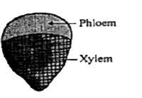
The tissue shown in the given figure is

AnguIar collenchyma, in which wall thickenings are present at the angles
Sclerenchyma, in which uniform wall thickenings are present
Xylem vessel that forms long channels for conduction of water and minerals
Phloem parenchyma with abundant food reserve.
The correct Answer is:C
Step by step video & image solution for The tissue shown in the given figure is by Biology experts to help you in doubts & scoring excellent marks in Class 9 exams.
Updated on:21/07/2023Topper's Solved these Questions
- TISSUES BOOK - SCIENCE OLYMPIAD FOUNDATION CHAPTER - TISSUES EXERCISE - ACHIEVERS SECTION (HOTS)
5 Videos
- NSO QUESTION PAPER 2020 SET 1BOOK - SCIENCE OLYMPIAD FOUNDATION CHAPTER - NSO QUESTION PAPER 2020 SET 1EXERCISE - ACHIEVERS SECTION
1 Videos
- WHY DO WE FALL ILLBOOK - SCIENCE OLYMPIAD FOUNDATION CHAPTER - WHY DO WE FALL ILLEXERCISE - MULTIPLE CHOICE QUESTIONS (ACHIEVERS SECTION (HOTS))
5 Videos
Similar Questions
Knowledge Check
Which statement is incorrect for the tissue shown in the given figure?
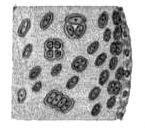 AThe tissue is usually covered by tough fibrous membrane called perichondrium.BThe cell embedded in the matrix of this tissue are called chondroblasts.CIt is found in nose tip, ear pinna, etc.DThe matrix is highly calcified and is richly supplied with nerves and blood vessels.
The value of the fraction, shown in the given figure, is
 A12B18C14D16
The value of the fraction, shown in the given figure, is
 A12B18C14D16
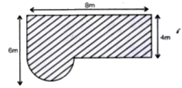
Find the area of the shaded field shown in the given figure
View SolutionIdentify the permanent tissues shown in the following figures .

View SolutionThe type of the vascular bundle shown in the given figure is
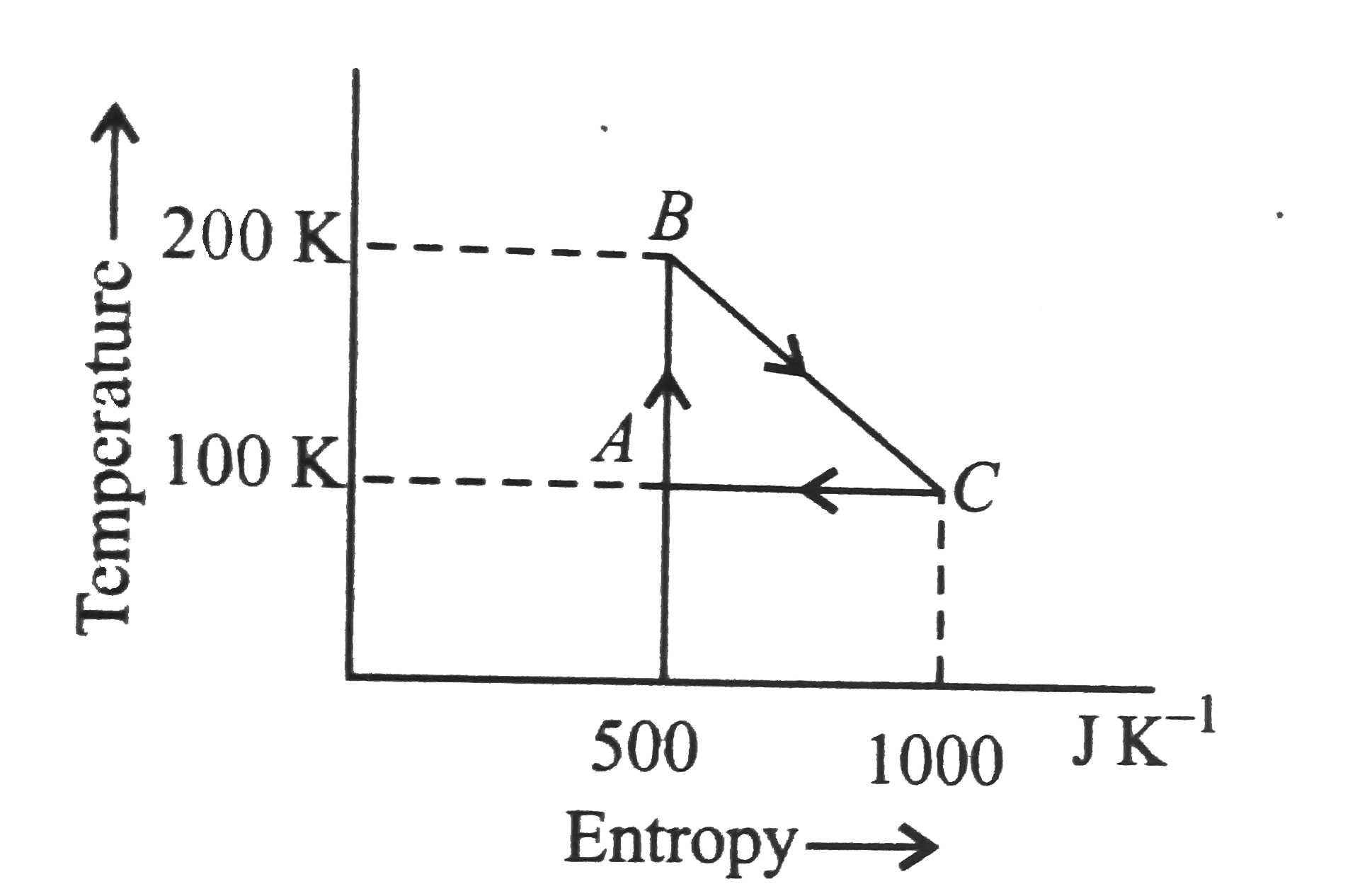
View SolutionThe efficiency of the reversible cycle shown in the given figure is
View SolutionThe value of fraction, shown in the given figure, is

View Solution

