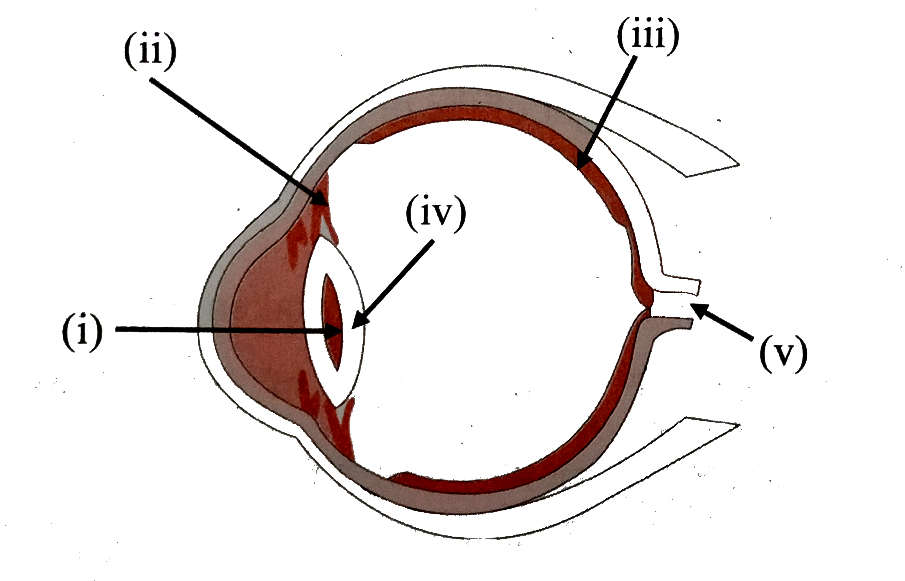
Label the parts of the following diagram of the human eye and state the functions of those parts.
Video Solution
Text Solution
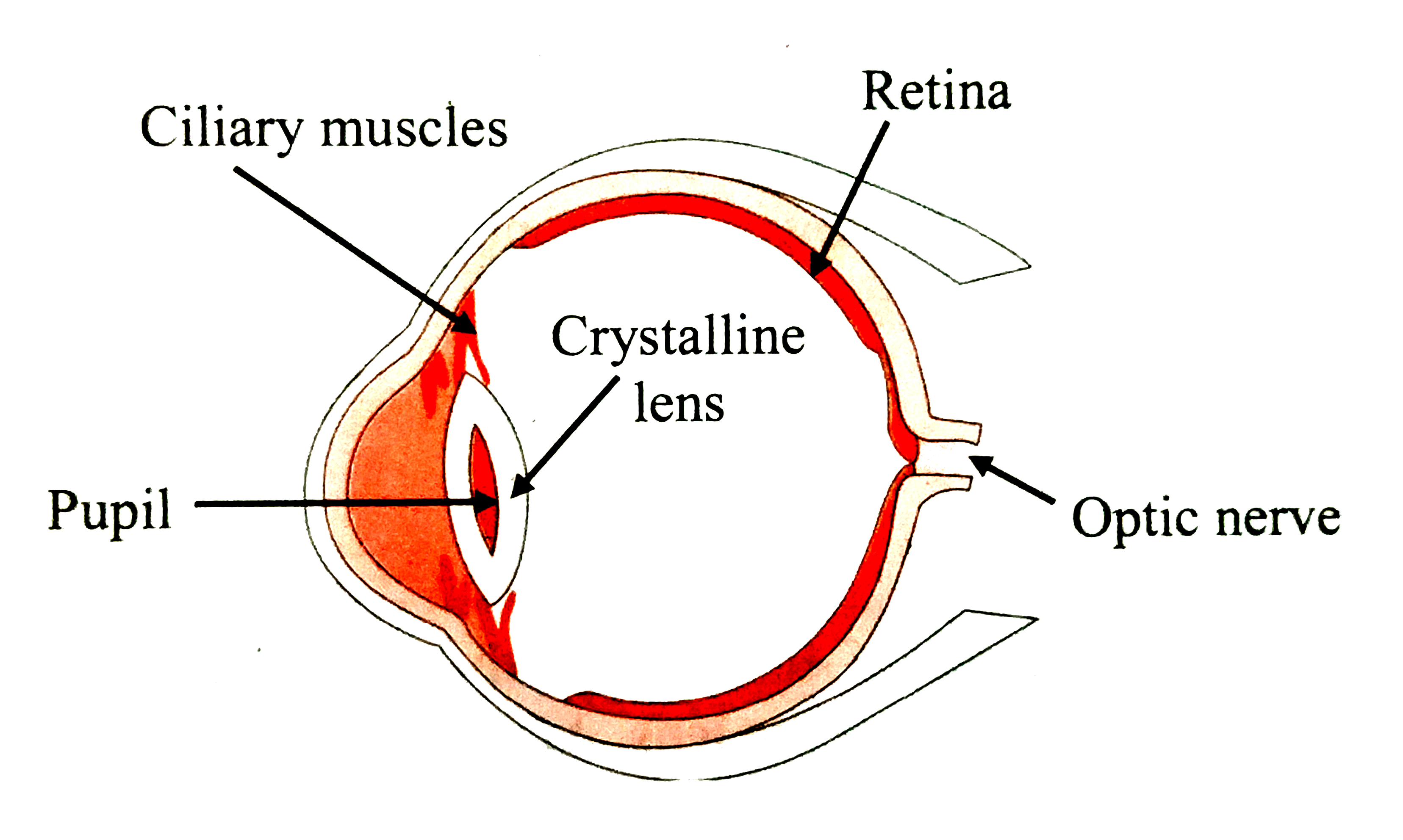
i. Pupil: The pupil contracts or widens depending on the intensity of light incident on the eye.
ii. Ciliary muscles: They help the eye lens to adjust the focal length depending on the distance of the object by contracting or relaxing.
iii. Retina: It is the screen inside the human eye which contains light sensitive cells. Image of an object is formed on this screen.
iv. Eye lens/crystalline lens: The lens allows the light to focus onto the retina.
v. Optic nerve: The sensitive cells on retina get excited when light falls on them and generate electric signals. Optic nerve carries these signals to the brain.
|
Topper's Solved these Questions
- LENSES BOOK - TARGET PUBLICATIONCHAPTER - LENSES EXERCISE - Questions based on paragraph
3 Videos
- LENSES BOOK - TARGET PUBLICATIONCHAPTER - LENSES EXERCISE - Practice problems
9 Videos
- LENSES BOOK - TARGET PUBLICATIONCHAPTER - LENSES EXERCISE - Complete the given chart/table
2 Videos
- HEATBOOK - TARGET PUBLICATIONCHAPTER - HEATEXERCISE - Values to remember
10 Videos
- MODEL QUESTION PAPER (PART -1 )BOOK - TARGET PUBLICATIONCHAPTER - MODEL QUESTION PAPER (PART -1 )EXERCISE - Questions
21 Videos
Similar Questions
Knowledge Check
The function of labelled part X is
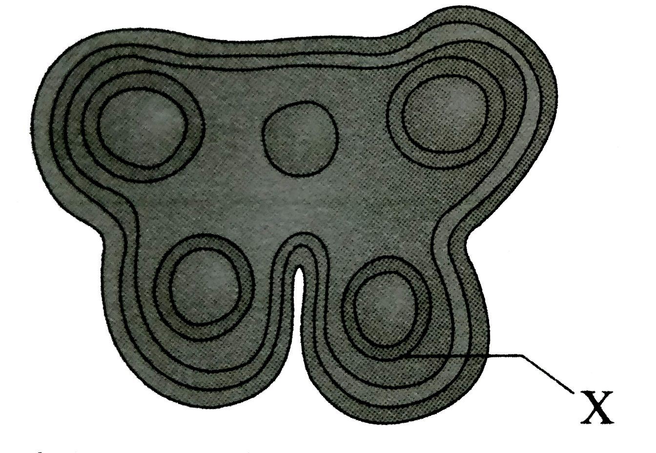 AdehiscenceBmechanicalCnutritionDprotection
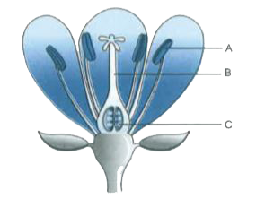
Name the parts A, B and C shown in the following diagram and state one function of each.
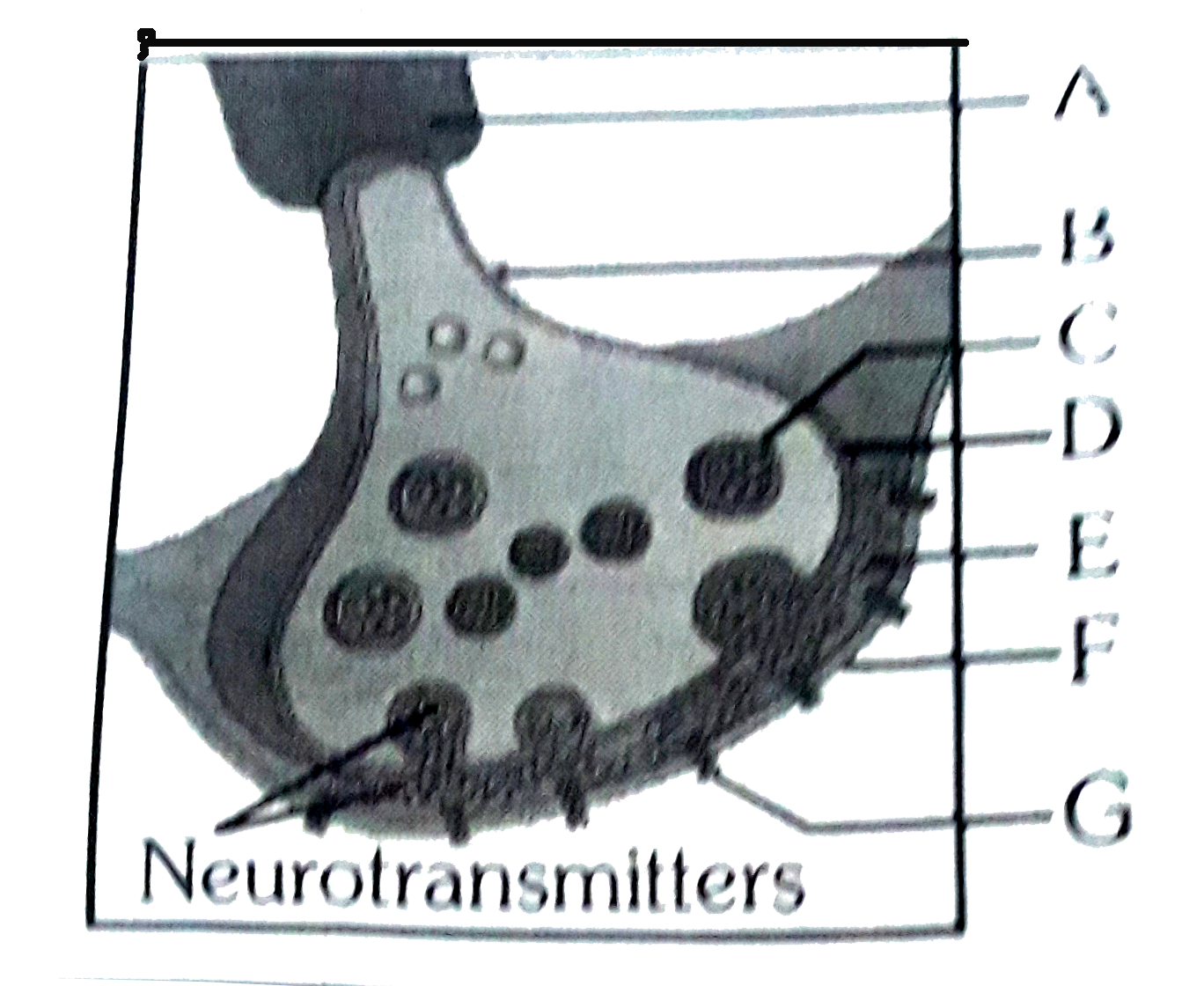
View SolutionLabel te parts in the given diagram of axon terminal and synapse
View SolutionDraw a diagram of the human excretory system and label the various parts.
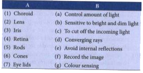
View SolutionMatch the parts of the human eye with its function .
View SolutionDraw a labelled sketch of the human eye. Explain the functions of each part.
View SolutionStudy the given diagram: Name the parts ‘A’ and ‘B’ and state one function of each.
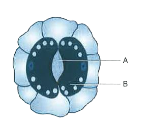
View SolutionStudy the given diagram: Name the parts ‘A’ and ‘B’ and state one function of each.

View Solution

