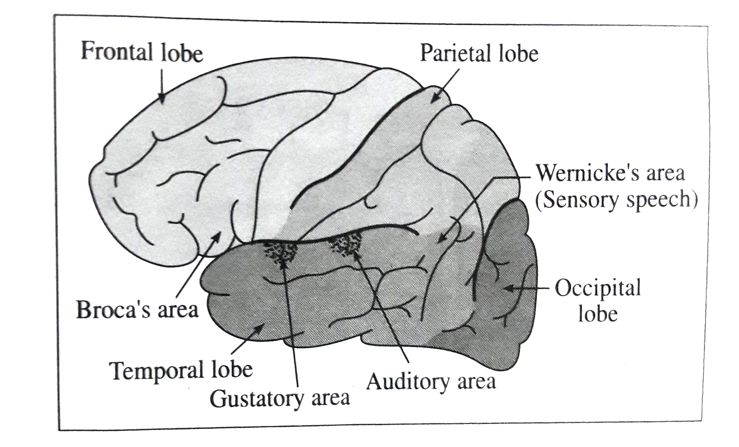
Each cerebral hemisphere is designed for specific function. Give details of these functions. Support your answer with a well labelled diagram .
1. Structure of cerebrum :
(1) Cerebrum is the largest part of the brain. It forms 80-85 % of the brain.
(2) A median longitudinal fissure divides the cerebrum into two cerebral hemispheres. These hemispheres are interspheres are interconnected by a thick band of transverse nerve fibres called corpus callosum.
(3) The outer part of cerebrum is called cortex while the inner part is called medulla.

(4) The roof of cerebrum is called pallium. Pallium is highly folded forming sulci and gyri. Sulci are depressions while gyri are ridges. The gyri increase the surface area of cerebal cortex.
(5) The ventrolateral walls of cerebrum are thickened and are called corpora striata.
(6) The cerebral cortex has three deep sulci, the central , lateral and parieto-oocipital.
(7) These sulci divide the cerebral hemisphere into four lobes. These are frontal, partietal, occipital and temporal lobes.
(8) The central sulcus separates frontal and parietal lobes. the lateral sulcus separates parietal and temporal lobes and the parieto-occipital sulcus separates parietal and occipital lobe.
(9) There are three functional areas in cerebral cortex. viz., sensory, association and motor area. Sensory area analyzes the sensory inputs. Association area processes the information .Motor area sends the motor outputs.
2. Functional areas of cerebrum are :
(1) General motor area and general sensory area.
(2) Association area
(3) Broca's area
(4) Sensory speech area or Wernick's area.
The functions given below are associated with these areas .
(6) Sensory visual area
(7) Auditory area.
3. Functions of cerebrum :
(1) The cerebrum controls the voluntary activities.
(2) The cerebrum perceives various sensory stimuli received through vision. taste , smell , sound , touch , speech , etc.
(3) It is the centre of memory, willpower , intelligence resoning and learning.
(4) The cerebrum is the centre for emotions, thoughts and fellings, pain , plessure, fear, fatigue, pressure, temperature , etc.
(5) It is also the centre for micturition, defecation , weeping , laughing etc.
Topper's Solved these Questions
- LONG ANSWER QUESTIONSBOOK - NAVNEET PUBLICATION - MAHARASHTRA BOARDCHAPTER - LONG ANSWER QUESTIONSEXERCISE - Human Reproduction
8 Videos
- LONG ANSWER QUESTIONSBOOK - NAVNEET PUBLICATION - MAHARASHTRA BOARDCHAPTER - LONG ANSWER QUESTIONSEXERCISE - Additional Important Questions for Self-practice
1 Videos
- LONG ANSWER QUESTIONSBOOK - NAVNEET PUBLICATION - MAHARASHTRA BOARDCHAPTER - LONG ANSWER QUESTIONSEXERCISE - Excretion and Osmoregulation
2 Videos
- GIVE REASONS BOOK - NAVNEET PUBLICATION - MAHARASHTRA BOARDCHAPTER - GIVE REASONS EXERCISE - Organisms and Environment - II
2 Videos
- MATCH THE COLUMNS/MATCH THE PAIRS/COMPLETE THE TABLEBOOK - NAVNEET PUBLICATION - MAHARASHTRA BOARDCHAPTER - MATCH THE COLUMNS/MATCH THE PAIRS/COMPLETE THE TABLEEXERCISE - Chapter 19 : Organisms and Environment - II
3 Videos
Similar Questions
Knowledge Check
Which lobe of cerebral hemisphere perform voluntary motor function the body?
AParietal lobeBFrontal lobeCOccipital lobeDTemporal lobe
Give one example to support your answer.
View SolutionExplain megasporogenesis till the formation of embryo sac. Support your answer with a labelled diagram.
View SolutionGive a brief account of human male reproductive system with well labelled diagrams.
View SolutionUnder what conditions a permanent electormagnet is obtained if a current carrying solenoid is used? Support your answer with the help of a labelled circuit diagram.
View SolutionUnder what conditions a permanent electormagnet is obtained if a current carrying solenoid is used? Support your answer with the help of a labelled circuit diagram.
View SolutionDescribe the structure and function of different type of epithelial tissue. Draw well labelled diagram.
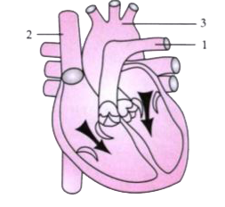
View SolutionThe diagram given below represents the human heart in one phase of its functional activities. Study the same and answer the questions that follow :

Draw well labelled diagrams of part 1 and 2 to show the structural differences between them.
View Solution
