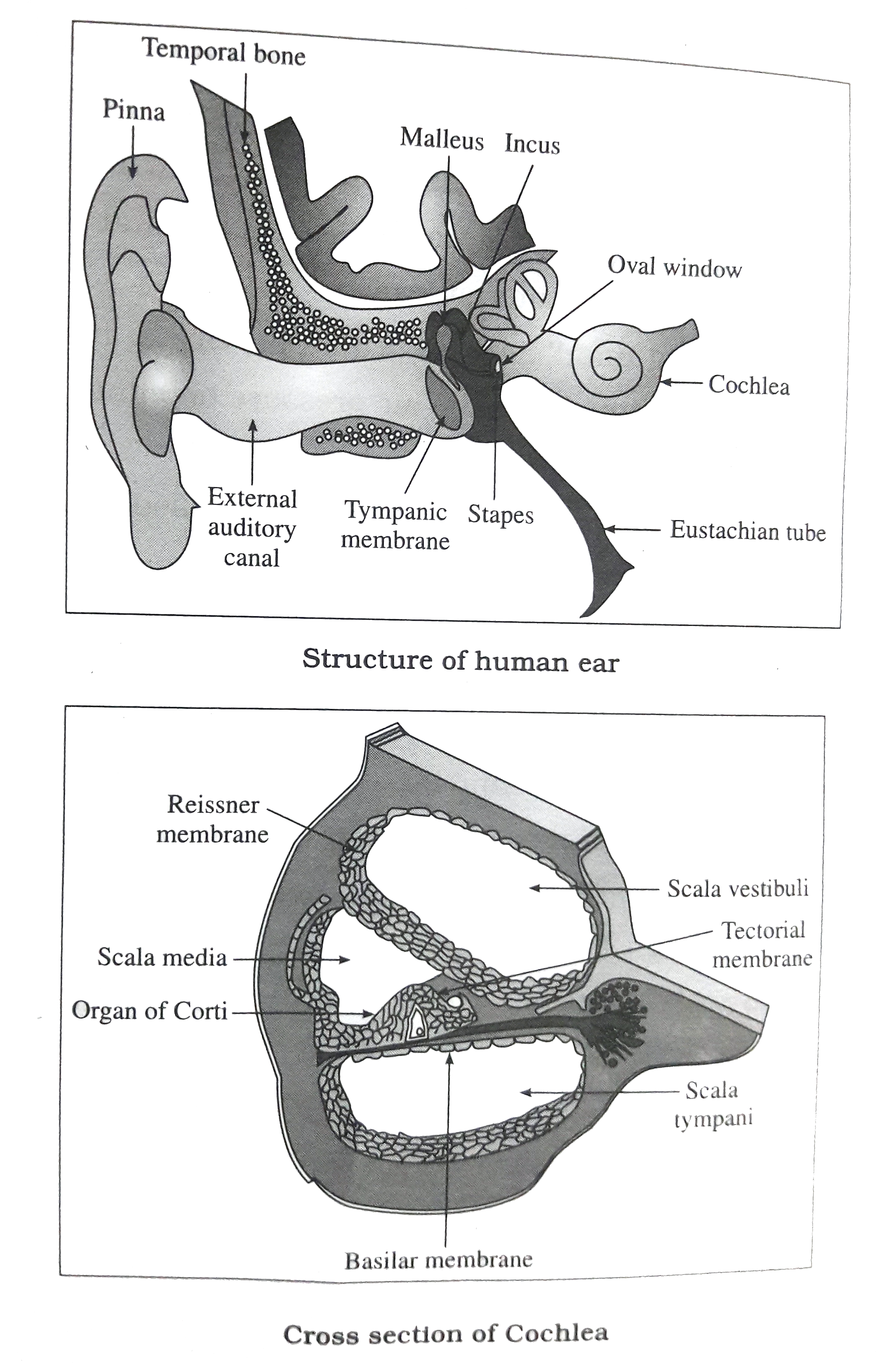
Briefly describe the structure of ear with the help of a suitable diagram.
Text Solution
(1) The ear is composed of the outer ear, middle ear and internal ear.
(2) The external ear consists of the pinna and external auditory meatus (canal). The pinna collects sound waves coming from the environment. The external auditory canal is the circular tube leading inside up to the ear drum. (tympanic membrane).
(3) The ear drum is formed of connective tissues having outer skin cover and inner mucus membrane.
(4) The middle ear consists of chain of three ossicles called malleus, incus and stapes. The malleusis attached to tympanic membrane and the stapes is connected to the oval window of the internal ear. They help in the transmission of sound waves from external auditory canal to internal ear.
(5) Connecting middle ear with the pharynx is an eustachian tube which helps in equalizing the air pressure on either side of the tympanic membrane.
(6) The internal ear is fluid filled structure called bony labyrinth and the membranous labyrinth.
(7) The membranes consist of coiled cochlea, the reissner's membrane and basilar membranes. These membranes divide the surrounding perilymph filled bony labyrinth into an upper scala vestibule and a lower scala tympani.
(8)The space within cochlea which is knwon as scala media is filled with endolymph. The scala vestibule ends at the oval window at the base of cochlea.
(9) The scala tympani terminates at the round window which opens to the middle ear. The organ of the corti is located on the brasillar membrane. It contains the hair cells which act as auditory receptors. A thin elastic membrane projects above the rows of the hairs cells called tectorial membrane.
(10) Above the cochlea. the internal ear also contains vestibular apparatus. It consists of three semicircular canals whcih are in perilymph and the otolith organs formed of the sacculus and utriculus.
(11) The bases of canals are swollen and are called ampullae.
Which contain a projecting ridge know as crista ampullaris which contain hair calls.
(12) The sacculus and utriculus also have projecting ridge called macula. The crista and macula are the specific receptors of vestibular apparatus . They are responsible for maintenance of body posture and the balance.
 .
.
|
Topper's Solved these Questions
- LONG ANSWER QUESTIONSBOOK - NAVNEET PUBLICATION - MAHARASHTRA BOARDCHAPTER - LONG ANSWER QUESTIONSEXERCISE - Human Reproduction
8 Videos
- LONG ANSWER QUESTIONSBOOK - NAVNEET PUBLICATION - MAHARASHTRA BOARDCHAPTER - LONG ANSWER QUESTIONSEXERCISE - Additional Important Questions for Self-practice
1 Videos
- LONG ANSWER QUESTIONSBOOK - NAVNEET PUBLICATION - MAHARASHTRA BOARDCHAPTER - LONG ANSWER QUESTIONSEXERCISE - Excretion and Osmoregulation
2 Videos
- GIVE REASONS BOOK - NAVNEET PUBLICATION - MAHARASHTRA BOARDCHAPTER - GIVE REASONS EXERCISE - Organisms and Environment - II
2 Videos
- MATCH THE COLUMNS/MATCH THE PAIRS/COMPLETE THE TABLEBOOK - NAVNEET PUBLICATION - MAHARASHTRA BOARDCHAPTER - MATCH THE COLUMNS/MATCH THE PAIRS/COMPLETE THE TABLEEXERCISE - Chapter 19 : Organisms and Environment - II
3 Videos
Similar Questions
Describe the structure of a sacromere with the help of a labelled diagram.
View SolutionDescribe the structure of a human kidney with the help of a labelled diagram.
View SolutionDescribe the structure of a human kidney with the help of a labelled diagram.
View SolutionDescribe the structure of anatropus ovule with the help of labelled diagram.
View SolutionBriefly describe the structure of the neuron.
View SolutionDescribe the structure of sex chromosomes with the help of labelled diagram.
View SolutionDescribe the internal ear of man with the help of a labelled diagram .
View SolutionBriefly describe the structure of the following:
Ear
View SolutionDescribe the structure of a human kidney with the help of a labelled diagram.
View SolutionDescribe the structure of a nucleosome with the help of a well-labelled diagram.
View Solution
