A plasmid DNA and linear DNA (both are of the same size) have one site for restriction endonuclease. When cut and separated on agarose gel electrophoresis, plasmid shows one DNA band, while linear DNA shows two fragments Explain.
Step-by-Step Solution:
1. Understanding Plasmid and Linear DNA:
- Plasmid DNA is typically circular in shape, while linear DNA is straight and has terminal ends. Both types of DNA can be cut by restriction endonucleases at specific sites.
2. Role of Restriction Endonucleases:
- Restriction endonucleases are enzymes that cut DNA at specific sequences. In this case, both the plasmid and linear DNA have one site for the restriction endonuclease.
3. Cutting Plasmid DNA:
- When the circular plasmid DNA is cut at its single restriction site, it opens up to form a linear piece of DNA. However, since it was originally circular, this single cut results in one continuous strand of DNA.
4. Cutting Linear DNA:
- When the linear DNA is cut at its single restriction site, it results in two separate fragments. This is because the linear DNA has two ends, and cutting it at one site divides it into two distinct pieces.
5. Separation by Agarose Gel Electrophoresis:
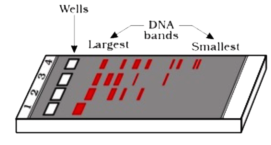
- When both types of DNA are subjected to agarose gel electrophoresis, the plasmid DNA, now a single linear strand, will migrate as one band. In contrast, the linear DNA will migrate as two separate bands due to the two fragments formed after the cut.
6. Conclusion:
- Therefore, the plasmid DNA shows one band on the gel, while the linear DNA shows two bands, reflecting the difference in the number of fragments produced after cutting by the restriction endonuclease.
- Plasmid DNA is typically circular in shape, while linear DNA is straight and has terminal ends. Both types of DNA can be cut by restriction endonucleases at specific sites.
- Restriction endonucleases are enzymes that cut DNA at specific sequences. In this case, both the plasmid and linear DNA have one site for the restriction endonuclease.
- When the circular plasmid DNA is cut at its single restriction site, it opens up to form a linear piece of DNA. However, since it was originally circular, this single cut results in one continuous strand of DNA.
- When the linear DNA is cut at its single restriction site, it results in two separate fragments. This is because the linear DNA has two ends, and cutting it at one site divides it into two distinct pieces.
- When both types of DNA are subjected to agarose gel electrophoresis, the plasmid DNA, now a single linear strand, will migrate as one band. In contrast, the linear DNA will migrate as two separate bands due to the two fragments formed after the cut.
- Therefore, the plasmid DNA shows one band on the gel, while the linear DNA shows two bands, reflecting the difference in the number of fragments produced after cutting by the restriction endonuclease.
Topper's Solved these Questions
Similar Questions
A plasmid DNA and a linear DNA (both are of the same size) have one site for a restriction endonuclease. When cut and separated on agarose gel electrophoresis, plasmid shows one DNA band while linear DNA shows two fragments. Explain
View SolutionA foreign DNA and plasmid cut by the same restriction endonuclease can be joined to form a recombinant plasmid using
View SolutionA foreign DNA and plasmid cut by the same restriction endonuclease can be joined to form a recombinant plasmid using
View SolutionGiven diagram showing a typical agarose gel electrophoresis showing migration of DNA fragments . The undigested DNA fragment will be present in the lane
 View Solution
With respect to RFLP , arrange the following sentences in order .
I. Agarose gel electrophoresis
II. Isolation on DNA
III . Cutting with Restriction Enzyme
IV. PCRView SolutionThe cutting of DNA by restriction endonucleases results in the fragments of DNA. These fragments can be separated by a technique known as
View SolutionThe cutting of DNA by restriction endonucleases results in fragments of DNA. These fragments are generally separated by a technique known as
View SolutionRead the following statements.
Statement A: In agarose gel electrophoresis, the smaller the fragment size of DNA, the farther it moves from the wall.
Statement B: Separation of proteins in normal electrophoresis is based on both size and charge.
Choose the correct option.View SolutionThis segment of DNA has restriction site-I and II, which create restriction fragments a, b and c. Which of the following gel (s) produced by electorphoresis would represent the separation and identification of these fragments ?
 View Solution
In the year 1963, the two enzymes responsible for restricting the growth of bacteriophage in E. coli were isolated. One of these added methyl groups to DNA, while the other cut DNA. The latter was called
View Solution

