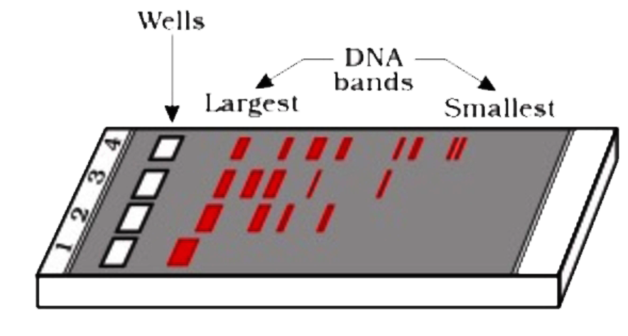
A plasmid DNA and a linear DNA (both are of the same size) have one site for a restriction endonuclease. When cut and separated on agarose gel electrophoresis, plasmid shows one DNA band while linear DNA shows two fragments. Explain
Step-by-Step Solution:
1. Understanding the Structure of DNA:
- Plasmid DNA is a circular DNA molecule, while linear DNA is a straight, linear structure. This fundamental difference in structure is crucial for understanding how they behave when cut by restriction endonucleases.
2. Cutting with Restriction Endonuclease:
- Both the plasmid DNA and the linear DNA have one site for a restriction endonuclease. When the restriction enzyme cuts the plasmid DNA, it opens the circular structure, resulting in one linear fragment of DNA.
- In contrast, when the linear DNA is cut at its restriction site, it will split into two separate linear fragments.
3. Separation by Agarose Gel Electrophoresis:
- When both types of DNA are subjected to agarose gel electrophoresis, they migrate through the gel based on their size and shape.
- The plasmid DNA, now in a linear form after being cut, will appear as one band on the gel because it is a single fragment.
- The linear DNA, having been cut into two fragments, will show two distinct bands on the gel.
4. Conclusion:
- Therefore, the plasmid DNA shows one band while the linear DNA shows two bands due to the difference in the number of fragments produced after the restriction enzyme cuts each type of DNA.
- Plasmid DNA is a circular DNA molecule, while linear DNA is a straight, linear structure. This fundamental difference in structure is crucial for understanding how they behave when cut by restriction endonucleases.
- Both the plasmid DNA and the linear DNA have one site for a restriction endonuclease. When the restriction enzyme cuts the plasmid DNA, it opens the circular structure, resulting in one linear fragment of DNA.
- In contrast, when the linear DNA is cut at its restriction site, it will split into two separate linear fragments.
- When both types of DNA are subjected to agarose gel electrophoresis, they migrate through the gel based on their size and shape.
- The plasmid DNA, now in a linear form after being cut, will appear as one band on the gel because it is a single fragment.
- The linear DNA, having been cut into two fragments, will show two distinct bands on the gel.
- Therefore, the plasmid DNA shows one band while the linear DNA shows two bands due to the difference in the number of fragments produced after the restriction enzyme cuts each type of DNA.
Topper's Solved these Questions
- BIOTECHNOLOGY : PRINCIPLES AND PROCESSESBOOK - ICSECHAPTER - BIOTECHNOLOGY : PRINCIPLES AND PROCESSESEXERCISE - PRACTICE QUESTIONS (VERY SHORT ANSWER TYPE QUESTIONS)
16 Videos
- BIOTECHNOLOGY : PRINCIPLES AND PROCESSESBOOK - ICSECHAPTER - BIOTECHNOLOGY : PRINCIPLES AND PROCESSESEXERCISE - PRACTICE QUESTIONS (SHORT ANSWER TYPE-I QUESTIONS)
13 Videos
- BIOLOGY-2016BOOK - ICSECHAPTER - BIOLOGY-2016EXERCISE - PART-II ( SECTION-C)
9 Videos
- BIOTECHNOLOGY AND ITS APPLICATIONS BOOK - ICSECHAPTER - BIOTECHNOLOGY AND ITS APPLICATIONS EXERCISE - COMPETITION CORNER (OBJECTIVE TYPE QUESTIONS (ASSERTION AND REASON TYPE QUESTIONS))
9 Videos
Similar Questions
A plasmid DNA and linear DNA (both are of the same size) have one site for restriction endonuclease. When cut and separated on agarose gel electrophoresis, plasmid shows one DNA band, while linear DNA shows two fragments Explain.
View SolutionA foreign DNA and plasmid cut by the same restriction endonuclease can be joined to form a recombinant plasmid using
View SolutionA foreign DNA and plasmid cut by the same restriction endonuclease can be joined to form a recombinant plasmid using
View SolutionGiven diagram showing a typical agarose gel electrophoresis showing migration of DNA fragments . The undigested DNA fragment will be present in the lane

View SolutionThe cutting of DNA by restriction endonucleases results in the fragments of DNA. These fragments can be separated by a technique known as
View SolutionThe cutting of DNA by restriction endonucleases results in fragments of DNA. These fragments are generally separated by a technique known as
View SolutionRead the following statements.
Statement A: In agarose gel electrophoresis, the smaller the fragment size of DNA, the farther it moves from the wall.
Statement B: Separation of proteins in normal electrophoresis is based on both size and charge.
Choose the correct option.
View SolutionWith respect to RFLP , arrange the following sentences in order .
I. Agarose gel electrophoresis
II. Isolation on DNA
III . Cutting with Restriction Enzyme
IV. PCR
View SolutionA DNA fragement was cleaved by using REN and the restriction fragments ( a,b and c ) produced as a result of digestion are shown below. Select the option that represents the correct gel electrophoresis separation of these fragments.

View SolutionIn the year 1963, the two enzymes responsible for restricting the growth of bacteriophage in E. coli were isolated. One of these added methyl groups to DNA, while the other cut DNA. The latter was called
View Solution
