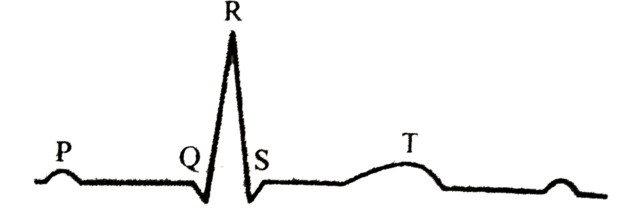
ECG depicts the depolarisation and repolarisation process during the cardiac cycle. In the ECG of a normal healthy individual one of the following waves is not represented.
Depolarisation of atria
Repolarisation of atria
Depolarisation of ventricles
Repolarisation of ventricles
The correct Answer is:C
Step-by-Step Solution:
1. Understanding ECG:
- ECG stands for electrocardiography, which is a method used to record the electrical activity of the heart over a period of time.
- It produces a graph that displays the electrical impulses in the heart.
2. Components of ECG:
- The ECG waveform consists of several key components: P wave, QRS complex, and T wave.
- Each of these waves represents different phases of the cardiac cycle.
3. P Wave:
- The P wave represents the depolarization of the atrial muscles. This is when the atria contract to push blood into the ventricles.
4. QRS Complex:
- The QRS complex is the most prominent part of the ECG and represents the depolarization of the ventricles. This is when the ventricles contract to pump blood to the lungs and the rest of the body.
- It also includes the repolarization of the atria, but this is not visually represented on the ECG.
5. T Wave:
- The T wave represents the repolarization of the ventricles. After the ventricles contract, they need to reset their electrical state to prepare for the next heartbeat.
6. Identifying the Missing Wave:
- In a normal ECG, the repolarization of the atria is not represented. While the depolarization of the atria (P wave) is shown, the repolarization of the atria occurs during the QRS complex but is not distinctly visible.
7. Conclusion:
- Therefore, in the ECG of a normal healthy individual, the wave that is not represented is the repolarization of the atria.
- ECG stands for electrocardiography, which is a method used to record the electrical activity of the heart over a period of time.
- It produces a graph that displays the electrical impulses in the heart.
- The ECG waveform consists of several key components: P wave, QRS complex, and T wave.
- Each of these waves represents different phases of the cardiac cycle.
- The P wave represents the depolarization of the atrial muscles. This is when the atria contract to push blood into the ventricles.
- The QRS complex is the most prominent part of the ECG and represents the depolarization of the ventricles. This is when the ventricles contract to pump blood to the lungs and the rest of the body.
- It also includes the repolarization of the atria, but this is not visually represented on the ECG.
- The T wave represents the repolarization of the ventricles. After the ventricles contract, they need to reset their electrical state to prepare for the next heartbeat.
- In a normal ECG, the repolarization of the atria is not represented. While the depolarization of the atria (P wave) is shown, the repolarization of the atria occurs during the QRS complex but is not distinctly visible.
- Therefore, in the ECG of a normal healthy individual, the wave that is not represented is the repolarization of the atria.
Topper's Solved these Questions
- BOOK - NCERT FINGERTPS ENGLISHCHAPTER - BODY FLUIDS AND CIRCULATIONEXERCISE - CORNER1videos
BODY FLUIDS AND CIRCULATION
- BOOK - NCERT FINGERTPS ENGLISHCHAPTER - BODY FLUIDS AND CIRCULATIONEXERCISE - Body Fluids And Circulation151videos
BODY FLUIDS AND CIRCULATION
- BOOK - NCERT FINGERTPS ENGLISHCHAPTER - BODY FLUIDS AND CIRCULATIONEXERCISE - HOTS5videos
BODY FLUIDS AND CIRCULATION
- BOOK - NCERT FINGERTPS ENGLISHCHAPTER - BIOMOLECULESEXERCISE - Biomolecules137videos
BIOMOLECULES
- BOOK - NCERT FINGERTPS ENGLISHCHAPTER - BREATHING AND EXCHANGE OF GASESEXERCISE - Breathing And Exchange Of Gases140videos
BREATHING AND EXCHANGE OF GASES
Similar Questions
Knowledge Check
- Question 1 - Select One
Which among the following is correct during each cardiac cycle ?
AThe volume of blood pumped out by the Rt (Right) and Lt (Left) ventricles is sameBThe volume of blood pumped out by the Rt and Lt ventricles is differentCThe volume of blood received by each atrium is differentDThe volume of blood received by the aorta and pulmonary artery is different - Question 2 - Select One
The given figure is the ECG of a normal human. Which one of its components is correctly interpreted below ?
 AComplex ORS - one complete pulseBPeak T - initiation of total cardiac contractionCPeak P and peak R together - systolic and diastolic blood pressuresDPeak P- initiation of left atrial contraction only
Explain the changes in heart during one cardiac cycle.
View SolutionWhich of the following is not represented in normal human ECG?
View SolutionThe tallest positive wave in ECG represents
View SolutionWhich among the following is correct during each cardiac cycle ?
View SolutionWhich among the following is correct during each cardiac cycle ?
View SolutionThe given figure is the ECG of a normal human. Which one of its components is correctly interpreted below ?
 View Solution

