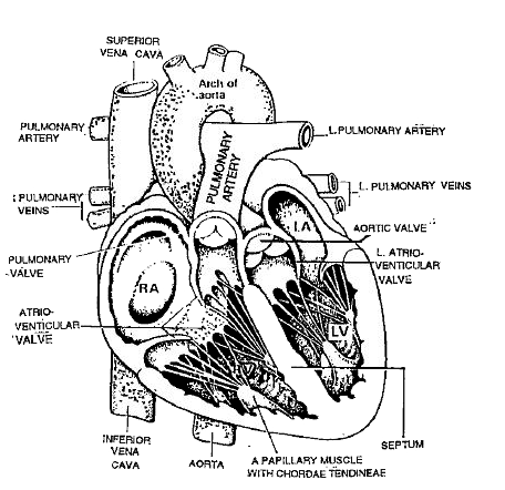
Describe the internal structure of heart of human beings.
Text Solution
Internal structure of human heart :

Auricles : Both auricles are thin walled because they have to push the blood only upto ventricles. Both auricles are smooth walled and have very low ridges called musculi pectinati. Both atria are separated by a thin vertical interauricular septum which prevents mixing of oxygenated and deoxygenated blood. A depression called fossa ovalis is present in interauricular septum which is a vestige of foramen ovale, present in the foetus.
Right auricle : It is larger in size than left auricle. It receives deoxygenated blood by superior vena cava (precaval) from upper portion of chest, inferior vena cava (post caval) from lower parts of body and coronary sinus from the wall of heart. Eustachian valve guards the opening of inferior vena cava and thebesian valve guards opening of coronary sinus. Right auricle opens into right ventricle via right auriculo-ventricular aperture which has a tricuspid valve, "Tricuspid valve prevents the back flow of blood from ventricle to auricle.
Left auricle : It receives oxygenated blood from lungs by four pulmonary veins. These have no valves. Left auricle opens into left ventricle by a crescentric aperture called left auriculoventricular aperture. It is guarded by a bicuspid valve (mitral valve) which regulates blood from left auricle to left ventricle but checks the backflow of blood.
Ventricles : The walls of ventricles are thicker than auricles. The walls of left ventricle is thicker than right ventricle because it has to pump the blood to all the parts of the body and right ventricle has to supply the blood only to lungs which are lying near the heart. The walls of ventricles have ridges and depression called columnae carneae and fissures respectively. These increase the surface area. The walls of ventricles have conical muscles called papillary muscles. Both ventricles are separated by a thick vertical interventricular septum which is obliquely placed. The septum has muscles called Bundle of His which gets branched and enter into walls of ventricles. The branches further get branched and the final branches are called Purkinje fibres. Pulmonary arch arises from left side at anterior end of right ventricle. It has three semilunar valves at its beginning. The pulmonary aorta divides into two pulmonary arteries which supply deoxygenated blood to lungs.
The systemic aorta arises from right side of left ventricle at anterior end. It also has three semilunar valves at its beginning. The systemic aorta is connected to pulmonary aorta by ligamentum arteriosum.
|
Similar Questions
Describe the internal structure of human ear.
View SolutionDescribe the internal structure of kidney.
View SolutionWith the help of a labelled diagram describe the internal structure of human heart.
View SolutionDescribe internal structure of a human heart.
View SolutionDescribe internal structure of a human heart.
View SolutionWith the help of well labelled diagram describe the internal structure of human heart .
View SolutionDraw the internal structure of human heart.
View SolutionDescribe the structure of the heart wall.
View SolutionDescribe the internal structure of human heart with the help of a diagram. Show the flow of blood with arrows.
View SolutionDescribe the structure of human heart.
View Solution
