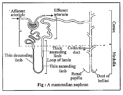
Describe the various parts of nephron with well Label diagram.
Text Solution
Each nephron is made up of two main parts :
I) Malpighian body or renal corpuscle : It comprises of two parts.
i) Bowman.s capsule ii) Glomerulus.
i) Bowman.s capsule : It is double walled cup-shaped structure. It is present in the cortex region of kidney. The two walls of Bowman.s capsule are as follows:
a) Inner wall or visceral wall: It is made up of squamous epithelial cells. It also contains few modified special cells, podocytes. Podocytes has finger like projections, called pedicels or feet. With the help of feet, it is connected with basement membrane of glomerulus.
b) Outer wall (Parietal wall): It is made up of unspecialised squamous epithelial cells. These cells are thin and flattened. They line over the basement membrane.
ii) Glomerulus : The cavity of Bowman.s capsule contains a bunch of blood capillaries, called Glomerulus. The position of glomerulus is maintained by connective tissue. The wall of glomerulus is made up of single layer of endothelial cells.
II) Renal tubule : The Bowman.s capsule opens into the renal tubule. Renal tubule consists of three parts.
i) Proximal convoluted tubule (PCT): It is the first part of renal tubule. It lies in the cortex region. It is much coiled structure. It is made up of cuboidal epithelium. The cuboidal epithelium gets modified into cone shaped cells. The latter bears microvilli to increase the surface area. In these cells, number of mitochondria and glycogen granules are present. They provide energy for active absorption.

ii) Loop of Henle :
Proximal convoluted tubules opens in loop of Henle. This part of renal tubule lies in medulla of kidney. Loop of Henle is U-shaped structure. It consists of two limbs :
a) Descending limb and b) Ascending limb.
a) Descending limb: It lies at the anterior end. PCT opens into it. It is thin walled structure. It consists of squamous epithelium. It is present in the medulla of kidney. Its wall is permeable to water and salt.
b) Ascending limb : It is thick walled structure. It is made up of cuboidal epithelium. Ascending limb is present in medulla but re-enters the cortex: It is impermeable to water but permeable to NaCl.
iii) Distal convoluted tubule (DCT): It is present in cortex region. Distal convoluted tubule is highly coiled structure. It is lined by cuboidal, columnar and ciliated columnar epithelium. The terminal end of DCT is straight and called tubule. Many tubules of nephrons opens into a large duct, are called collecting duct.
Collecting duct : It is of large size. It lies in cortex as well as in medulla of kidney. Many collecting ducts further unite to form a large duct. It is known as Duct of Bellini. The wall of collecting duct is lined by cuboidal columnar as well as ciliated columnar epithelium. Duct of Bellini opens into the renal pyramid. From here, it goes into renal pelvis and then to ureter.
|
Similar Questions
Describe the parts of a fruit. Draw a well labelled diagram showing the different parts of a fruit.
View SolutionDescribe the process of fertilization with a well-labelled diagram.
View SolutionDescribe the double helix model of DNA with well labelled diagram
View SolutionExplain the structure of a nephron with the help of a labelled diagram.
View SolutionDescribe various types of epithelial tissues with the help of labelled diagrams.
View SolutionDescribe various types of epithelial tissues with the help of labelled diagrams.
View SolutionDescribe various types of epithelial tissues with the help of labelled diagrams.
View SolutionDescribe the process of filtration with the help of a well-labelled diagram.
View SolutionDescribe the structure of a nucleosome with the help of a well-labelled diagram.
View SolutionDraw a labelled diagram of a nephron.
View Solution
