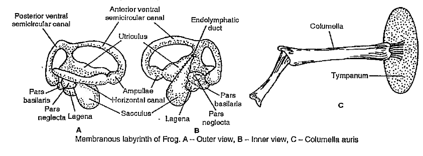
Describe structure of ear of frog.
Text Solution
Ear : It is the organ of hearing and equilibrium
Parts : It has middle and internal ear.
Middle ear : The cavity of middle ear is called tympanic cavity. It is closed externally by the e drum or tympanic membrane which is a dark circular patch of skin behind eye. It is tightly stretched over annulus tympanicus. Middle ear cavity contains air and communicates with buccopharyngea cavity by the eustachian tube. The bony partition between tympanic cavity and auditory capsule is perforated by oval operture the fenestra oval is, which remains closed by a membrane and a carilaginou nodule the stapedial plate. A rod of bone and cartilage the columella auris, extends across the tympani cavity. Its outer end is attached to the ear drum while inner end to the stapedial plate.
Internal ear : The internal ear or auditory capsule is formed by the prootic and exoccpital bones. Its cavity is filled with perilymph into which floats inner ear or membranous labyrinth. Its sac like body or vestibule is made of a larger utriculus and a smaller sacculus. Sacculus forms two small rounded diverticuala, lagena and pars basilaris. Lagena is the forerunner of the cochlear duct of higher vertebrates Pars basilaris seems to be a part oflagena, Above the two is another protuberance, pars neglecta, which

comes from utriculus. Three semicircular canals anterior and posterior vertical and horizontal-arise from utriculus and re arranged at right angles with one another. Only one end of each canal is slightly swollen into an ampulla. A small narrow endolymphatic duct arises from inner dorsal end of sacculus and terminates in an expanded thin walled endolymphatic sac over hind brain.
Histology of membranous labyrinth : Membranous labyrinth is filled with a milky fluid the enciolymph containing small particles of calcium carbonate, called cotoliths. The thin wall of labyrinth is cuboidal epithelium which is modified forming sensory. patches called acoustic spots or ridges. These form the actual receptors of ear. An acoustic ridge is made up of supporting cells and sensory hair cells.
The sensory spots of ampullae are called cristae and in vestibule are called maculae.
Working of ear : Sound waves strike tympanum and vibrates, these are transmitted to perilymph through columella and stepedial plate. From perilymph sound waves pass to endolymph of internal ear and stimulate sensory hair cells of Jagena. These are carried to brain.
Equilibrium : Utriculus and semicircular canals are concerned with equilibrium. Otoliths are disturbed by change in position of body. These otoliths stimulate hair and message is carried by auditory nerve to rain which directs body muscles to balance.
|
Updated on:21/07/2023
Similar Questions
Discuss structure of heart of frog.
View SolutionBriefly describe the structure of the following:
EarView SolutionBriefly describe the structure of ear with the help of a suitable diagram.
View SolutionBriefly describe the structure of the following :
(a) Brain
(b) Eye
(c) EarView SolutionDescribe the manus or pes of frog.
View SolutionStructure Of Human Ear
View SolutionName the parts of ear in frog.
View SolutionName the parts of ear in frog.
View SolutionDescribe the internal structure of human ear.
View SolutionBones of the hands, structure of nostrils, position of eyes, structure of ear pinnae
View Solution

