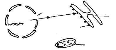
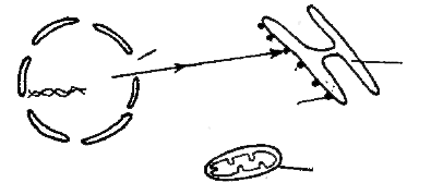
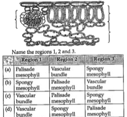
The diagram shows part of a leaf as seen in cross section under the microscope.
Text Solution
The correct Answer is:C
The vascular bundles in leaves are present in the veins of the leaf, which act as a transport medium (Region 1). Mesophyll is the internal ground tissue located between the two epidermal cell layers of the leaf, and is composed of two kinds of tissues: the palisade parenchyma, located beneath the upper epidermis (Region 2) and composed of columnar cells oriented perpendicular to the leaf surface, and spongy parenchyma, located in the lower part of the leaf and composed of irregularly shaped cells (Region 3).
|
Topper's Solved these Questions
- PHOTOSYNTHESISBOOK - ICSECHAPTER - PHOTOSYNTHESISEXERCISE - ASSERTION & REASON
6 Videos
- PHOTOSYNTHESISBOOK - ICSECHAPTER - PHOTOSYNTHESISEXERCISE - STATE THE FUNCTION OF THE FOLLOWING:
2 Videos
- NERVOUS SYSTEM BOOK - ICSECHAPTER - NERVOUS SYSTEM EXERCISE - REVIEW QUESTIONS (Find the odd one out: )
10 Videos
- POLLUTION BOOK - ICSECHAPTER - POLLUTION EXERCISE - REVIEW OLUESTIONS
55 Videos
Similar Questions
Knowledge Check
Minimum cell size seen under light microscope is
A1μmB0⋅1μmC0⋅25μmD0⋅5μmGolgi apparatus was first seen under electron microscope by
ANovikroffBDalton and FelixCRhodinDDe Robertis and Franchi
The diagram below-shows features of an animal cell as seen under electron microscope :
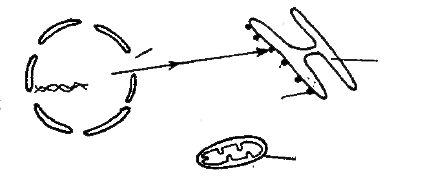
Name the structure B.
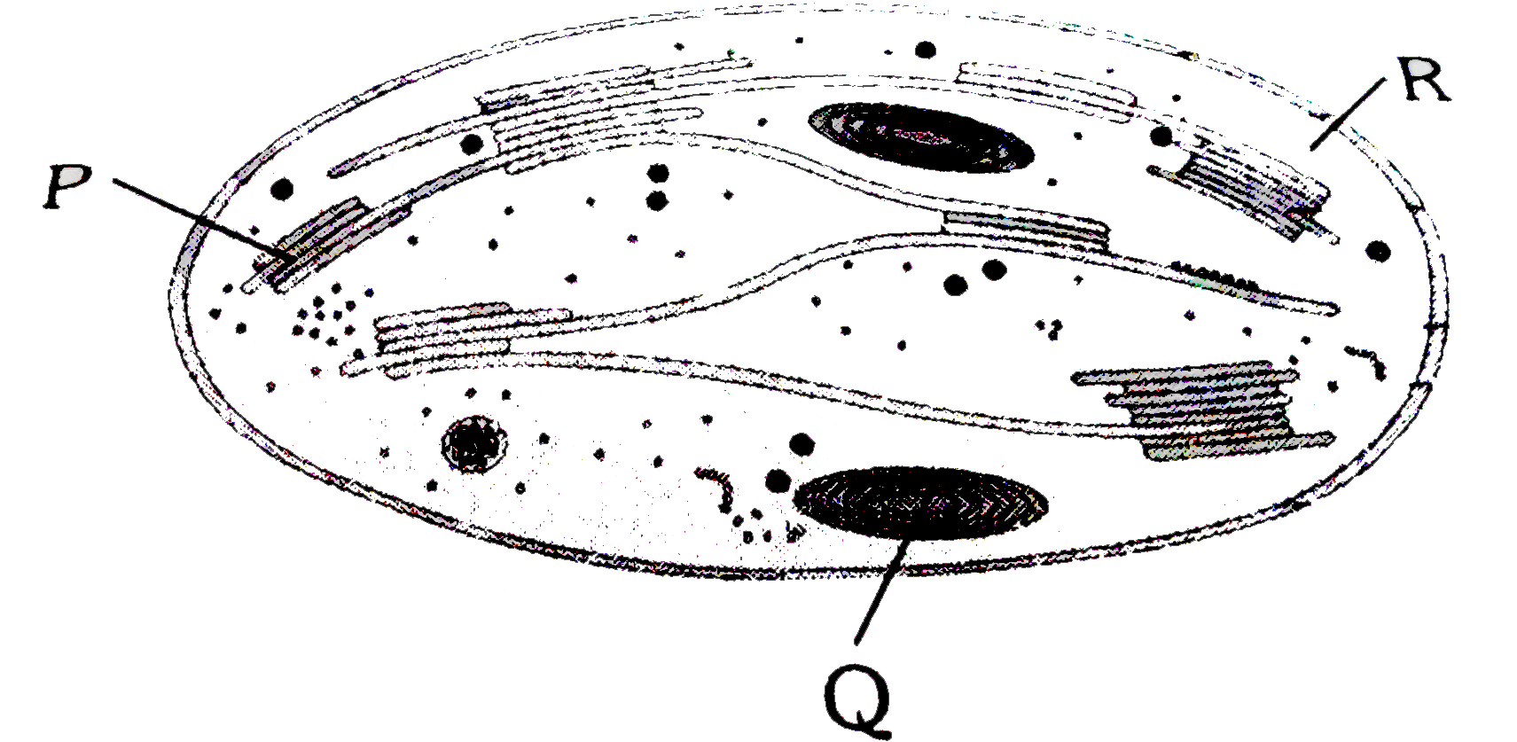
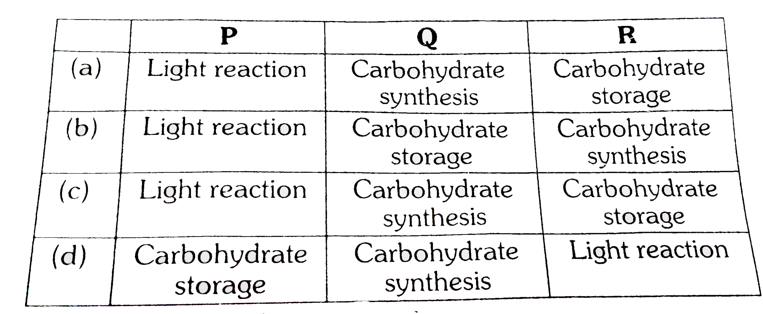
View SolutionThe diagram shows the ultra structure of a chloroplast as seen in section .Identify the functions of P,Q and R
View SolutionA student is observing the temporary mount of a leaf peel under a microscope. Draw labelled diagram of the structure (of stomata) as seen under the microscope.
View SolutionA student is observing the temporary mount of a leaf peel under a microscope. Draw
labelled diageram of the structure (of stomata) as seen under the microscope.
View SolutionMinimum cell size seen under light microscope is
View SolutionGolgi apparatus was first seen under electron microscope by
View Solution

