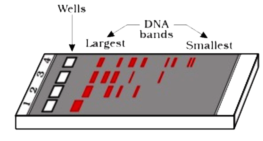
A plasmid DNA and a linear DNA both are of the same size have one site for a restriction endonuclease. When cut and separated on agarose gel electrophoresis plasmid shows one DNA band while linear DNA shows two fragments. Explain.
It is because plasmid is a circular DNA molecule. When cut with enzyme it becomes linear but does not get fragmented. Whereas a linear DNA molecule gets cut into two fragments. Hence a single DNA band is observed for plasmid while two DNA bands are observed for linear DNA in agarose gel.
Related Solutions
A plasmid DNA and a linear DNA (both are of the same size) have one site for a restriction endonuclease. When cut and separated on agarose gel electrophoresis, plasmid shows one DNA band while linear DNA shows two fragments. Explain.
View SolutionA plasmid DNA and linear DNA (both are of the same size) have one site for restriction endonuclease. When cut and separated on agarose gel electrophoresis, plasmid shows one DNA band, while linear DNA shows two fragments Explain.
View SolutionA plasmid DNA and a linear DNA (both of the same size) have one site for a restriction endonuclease. When cut and separated on agarose gel electrophoresis, plasmid shows one DNA band while linear DNA shows two fragments. Explain.
View SolutionA plasmid DNA and a linear DNA (both are of the same size) have one site for a restriction endonuclease. When cut and separated on agarose gel electrophoresis, plasmid shows one DNA band while linear DNA shows two fragments. Explain
View SolutionA plasmid DNA and a linear DNA (both of the same size) have one site for a restriction endonuclease, When cut and separated on agarose gel electrophoresis, plasmid shows one DNA band while, linear DNA shows two fragments. Explain.
View SolutionA plasmid DNA and linear DNA (both are of the same size) have one site for restriction endonuclease. When cut and separated on agarose gel electrophoresis, plasmid shows one DNA band, while linear DNA shows two fragments Explain.
View SolutionA plasmid DNA and a linear DNA , both are of the same size have one site for a restriction endonulease . When cut and separated on agarose gel electrophoresis,
View SolutionIn a agarose gel electrophoresis , the DNA fragments are separated according to their
View SolutionGiven diagram showing a typical agarose gel electrophoresis showing migration of DNA fragments . The undigested DNA fragment will be present in the lane
 View Solution
Given diagram showing a typical agarose gel electrophoresis showing migration of DNA fragments . The undigested DNA fragment will be present in the lane
 View Solution
Agarose gel electrophoresis separates DNA molecules of size.
View SolutionHow many linear DNA fragments will be produced when a circular plasmid is digested with a restriction enzyme having 3 sites?
View SolutionSeparation of DNA fragments using agarose gel electrophoresis occurs due to:
View SolutionA foreign DNA and plasmid cut by the same restriction endonuclease can be joined to form a recombinant DNA by :
View Solutionप्रक्कथन : पौधों के वर्णकों का मिश्रण क्रोमेटोग्राफी द्वारा पृथक किया जा सकता है। कारण : क्रोमेटोग्राफी रंगीन पदार्थो को अकेले संघटकों में पृथक करने के लिये उपयोगी है।
View SolutionProcesses OF rDNA Technology:- Introduction || Isolation OF DNA || Fragmentation OF DNA by RE || Separation and Isolation OF Desired DNA Fragment (Agarose Gel Electrophoresis)
View SolutionProcesses OF rDNA Technology:- Introduction, Isolation OF DNA, Fragmentation OF DNA by RE, Separation and Isolation OF Desired DNA Fragment (Agarose Gel Electrophoresis)
View Solutionऐगारोज जैल इलेक्ट्रोफोरेसिस में DNA अणु पृथक् होते हैं
View SolutionIn agarose gel electrophoresis, DNA molecules are separated on the basiS of their
View SolutionIn agarose gel electrophoresis, DNA molecules are separated on the basis of their :
View Solution
