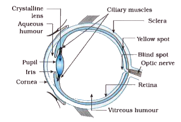
With a neat labelled diagram, explain the structure and functioning of the human eye.

These parts and their functions are as follows:
The human eyeball is approximately spherical in shape and its diameter is about 2.3 cm.
(1) Cornea : The front portion of the eye, bulging outwards, is known as the cornea.
It is a transparent and thin spherical membrane.
Light enters the eye through this membrane. Most of the refraction of light rays entering the eye occurs at the outer surface of the cornea, i.e., cornea refracts the rays of light falling on the eye.
(2) Iris : Beyond the cornea, there is a dark coloured muscular diaphragm known as the iris.
The iris controls the size of the pupil.
It is the colour of the iris that we call as the colour of the eye.
(3) Pupil : The small hole in the middle of the iris is called the pupil.
Light enters the eye through the pupil.
The pupil regulates and controls the amount of light entering into the eye.
[The pupil of the eye acts like a variable aperture whose size varies with the help of the iris.
When the light is very bright, the iris contracts the pupil to allow less light to enter into the eye.
However, in dim light the iris expands the pupil to allow more light to enter into the eye.
Thus, the pupil opens completely through relaxation of the iris.]
(4) Crystalline lens : Behind the iris there is the eye lens which is a convex lens made of a transparent, soft and flexible material like a jelly made of proteins.
The focal length and hence converging power of the eye lens is adjustable, because its curvature can be changed (modified) to some extent by ciliary muscles.
It focuses an inverted real image of the object on the retina.
(5) Ciliary muscles : They hold the eye lens in position and help in changing (modifying) the curvature of the eye lens and hence the focal length of the eye lens.
When the ciliary muscles are relaxed, the eye lens becomes thin. Hence, its focal length increases and the converging power decreases.
When the ciliary muscles are tense, the eye lens becomes thick. Hence, its focal length decreases and the converging power increases.
(6) Retina : Behind the eye lens and at the back side of the eyeball, there is a screen called retina on which an inverted real image of the object is formed.
The retina is a delicate membrane / film covering the entire curved back surface of the eye. It has enormous number of light-sensitive cells in the form of rods and cones.
The rod-like cells respond to the intensity of light and cone-like cells respond to the colour of light.
These cells get activated upon illumination and generate electrical signals (or nerve impulses) which are sent to the brain via the optic nerve.
The brain interprets these signals and finally, processes the information so that we perceive objects as they are.
(7) Optic nerve : It transmits visual information from the retina to the brain.
( 8 ) Aqueous humour: The space between the cornea and eye lens is filled with a viscous and transparent liquid called aqueous humour which helps the refracted light to be focussed on the retina. It also maintains intraocular pressure.
(9) Vitreous humour : The space between the eye lens and retina contains transparent jelly called the vitreous humour.
Topper's Solved these Questions
- THE HUMAN EYE AND THE COLOURFUL WORLDBOOK - KUMAR PRAKASHANCHAPTER - THE HUMAN EYE AND THE COLOURFUL WORLDEXERCISE - TEXTUAL EXERCISE
13 Videos
- THE HUMAN EYE AND THE COLOURFUL WORLDBOOK - KUMAR PRAKASHANCHAPTER - THE HUMAN EYE AND THE COLOURFUL WORLDEXERCISE - ADDITIONAL QUESTIONS AND ANSWERS
9 Videos
- QUESTION PAPER-2 (MARCH,2020 - BOARD'S QUESTIONS PAPER)BOOK - KUMAR PRAKASHANCHAPTER - QUESTION PAPER-2 (MARCH,2020 - BOARD'S QUESTIONS PAPER)EXERCISE - Section - D (Answer the following questions)
3 Videos
Similar Questions
Write the help of a well labelled diagram, explain the construction and working of the human eye .
View SolutionExplain the structural and functional significance of fovea in the human eye.
View SolutionDraw a neat and labelled diagram of the human ear. With the help of this diagram, explain the construction and working of the human ear.
View SolutionWith a neat labelled diagram, explain how a rainbow is formed.
View SolutionWith a neat labelled diagram of the structure, explain formation of a p-type semiconductor.
View SolutionDraw a neat and labelled diagram showing the structure of an atom.
View SolutionWith the hepl of a diagram, explain the structure of human eye.
View SolutionDraw a neat labelled diagram to show the structure of the human eye.
View SolutionWith the help of a neat and labelled diagram describe the anatomy of 'human eye'. Explain the mechanism of vision.
View SolutionWith the help of a neat and labelled diagram, describe anatomy of human eye. Explain the mechanism of vision .
View Solution
